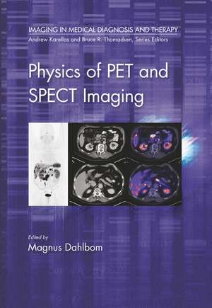
Physics of PET and SPECT Imaging: Imaging in Medical Diagnosis and Therapy
Editat de Magnus Dahlbomen Limba Engleză Paperback – 31 mar 2021
Din seria Imaging in Medical Diagnosis and Therapy
- 9%
 Preț: 572.72 lei
 - 5%
 Preț: 338.80 lei
 -
 Preț: 359.60 lei
 -
 Preț: 436.14 lei
 -
 Preț: 436.14 lei
 - 16%
 Preț: 302.11 lei
 -
 Preț: 436.14 lei
 - 12%
 Preț: 317.92 lei
 - 12%
 Preț: 325.34 lei
 - 10%
 Preț: 326.37 lei
 - 17%
 Preț: 296.80 lei
 - 11%
 Preț: 321.92 lei
 - 12%
 Preț: 317.92 lei
 - 15%
 Preț: 427.16 lei
 - 10%
 Preț: 326.09 lei
 - 30%
 Preț: 820.03 lei
 - 17%
 Preț: 296.80 lei
 - 5%
 Preț: 426.94 lei
 - 25%
 Preț: 610.27 lei
 - 15%
 Preț: 461.03 lei
 - 22%
 Preț: 477.32 lei
 - 12%
 Preț: 313.61 lei
 - 16%
 Preț: 299.92 lei
 - 12%
 Preț: 315.28 lei
 - 5%
 Preț: 1548.34 lei
 - 12%
 Preț: 317.18 lei
 - 21%
 Preț: 415.10 lei
 - 12%
 Preț: 314.03 lei
 - 12%
 Preț: 312.43 lei
 - 22%
 Preț: 434.76 lei
 - 11%
 Preț: 321.33 lei
 -
 Preț: 449.25 lei
 - 13%
 Preț: 322.04 lei
 - 18%
 Preț: 1117.07 lei
 - 17%
 Preț: 464.79 lei
 - 12%
 Preț: 317.58 lei
Preț: 325.33 lei
Preț vechi: 363.14 lei
-10% Nou
Puncte Express: 488
Preț estimativ în valută:
62.27€ • 64.75$ • 52.10£
62.27€ • 64.75$ • 52.10£
Carte tipărită la comandă
Livrare economică 14-28 martie
Preluare comenzi: 021 569.72.76
Specificații
ISBN-13: 9780367782368
ISBN-10: 0367782367
Pagini: 503
Dimensiuni: 178 x 254 mm
Greutate: 1.07 kg
Ediția:1
Editura: CRC Press
Colecția CRC Press
Seria Imaging in Medical Diagnosis and Therapy
ISBN-10: 0367782367
Pagini: 503
Dimensiuni: 178 x 254 mm
Greutate: 1.07 kg
Ediția:1
Editura: CRC Press
Colecția CRC Press
Seria Imaging in Medical Diagnosis and Therapy
Cuprins
Part 1 BASICS. Principles of SPECT and PET Imaging. Part 2 TECHNOLOGY. Scintillators for PET and SPECT. Photodetectors. Acquisition Electronics. SPECT Instrumentation. PET Instrumentation. PART 3 QUANTITATIVE IMAGING. Methodologies for Quantitative SPECT. Data Corrections and Quantitative PET. Image Reconstruction for PET and SPECT. High-Performance Computing in Emission Tomography. Methods and Applications of Dynamic. SPECT Imaging. Dynamic PET Imaging. Part 4 MULTIMODALITY IMAGING. PET/CT. SPECT/CT. PET/MRI. Part 5 PRECLINICAL IMAGING AND CLINICAL APPLICATIONS. Preclinical PET and SPECT. Clinical Applications of PET/CT and SPECT/CT Imaging
Notă biografică
Magnus Dahlbom has been working in the field of Nuclear Medicine for close to 30 years. He earned his B.Sc. in physics from the University of Stockholm in 1982. He received his Ph.D. from UCLA in 1987. His Ph.D. research was on high-resolution PET detectors and image processing. In 1989 he was part of the team that started the first clinical PET operation in the U.S. at UCLA. Around the same time developed together with Drs. Edward J. Hoffman and Michael E. Phelps the Whole Body PET imaging technique, which currently makes up more than 90% of all PET studies performed. His research interests are in PET and SPECT instrumentation and image processing. Since 1989 he has been the chief physicist at the Nuclear Medicine services at UCLA where he is responsible for all imaging instrumentation, including SPECT, SPECT/CT and PET/CT systems. At UCLA he is the faculty graduate advisor in the Biomedical Physics graduate program. He is teaching graduate level courses on the basics of Nuclear Medicine imaging and instrumentation. He has authored and co-authored more than 120 research papers and 11 book chapters on nuclear imaging instrumentation and processing. He was also co-editor of a PET/CT atlas. For the last 6 years he has been serving as an editorial consultant to the editor-in-chief of the Journal of Nuclear Medicine.
Recenzii
"A well-written, comprehensive, advanced-level book that also provides enough information about the physics, mathematics, instrumentation, quantification, and clinical applications of these imaging modalities."
-Jun Zhang, The Ohio State University
-Jun Zhang, The Ohio State University
Descriere
This text provides coverage of PET and SPECT instrumentation and multimodality imaging. It takes an integrative approach, bridging the researcher and clinician’s perspectives. It begins with an introduction to basic physics of PET and SPECT, followed by a section on detector technology. It addresses various aspects of producing
