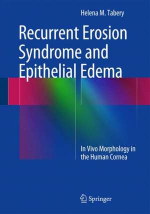
Recurrent Erosion Syndrome and Epithelial Edema: In Vivo Morphology in the Human Cornea
Autor Helena M. Taberyen Limba Engleză Hardback – 4 dec 2014
| Toate formatele și edițiile | Preț | Express |
|---|---|---|
| Paperback (1) | 702.49 lei 38-44 zile | |
| Springer International Publishing – 22 sep 2016 | 702.49 lei 38-44 zile | |
| Hardback (1) | 720.31 lei 22-36 zile | |
| Springer International Publishing – 4 dec 2014 | 720.31 lei 22-36 zile |
Preț: 720.31 lei
Preț vechi: 758.23 lei
-5% Nou
Puncte Express: 1080
Preț estimativ în valută:
137.83€ • 144.29$ • 114.05£
137.83€ • 144.29$ • 114.05£
Carte disponibilă
Livrare economică 17-31 martie
Preluare comenzi: 021 569.72.76
Specificații
ISBN-13: 9783319065441
ISBN-10: 3319065440
Pagini: 150
Ilustrații: XIII, 154 p. 150 illus. in color.
Dimensiuni: 178 x 254 x 17 mm
Greutate: 0.53 kg
Ediția:2015
Editura: Springer International Publishing
Colecția Springer
Locul publicării:Cham, Switzerland
ISBN-10: 3319065440
Pagini: 150
Ilustrații: XIII, 154 p. 150 illus. in color.
Dimensiuni: 178 x 254 x 17 mm
Greutate: 0.53 kg
Ediția:2015
Editura: Springer International Publishing
Colecția Springer
Locul publicării:Cham, Switzerland
Public țintă
Professional/practitionerCuprins
CORNEAL RECURRENT EROSION SYNDROME: The Morphology of Recurrent Erosions.- Case Reports.- Recurrent Erosions and Stromal Infiltrates. CORNEAL EPITHELIAL EDEMA: The Morphology of Epithelial Edema.- Case Reports. Final Remarks.
Notă biografică
Helena M. Tabery gained her MD from the University of Lund, Sweden in 1972 and thereafter undertook ophthalmologic training at the Eye Clinic, Malmö University Hospital UMAS, Sweden (1973-1975) and the Eye Department of Doc. Dr. Karl Lisch in Wörgl, Austria (1975-77). Dr. Tabery has been an accredited Specialist in Ophthalmology since 1975. Between 1977 and 1989 she was a clinical teacher at the Department of Ophthalmology, Malmö University Hospital, University of Lund, Malmö, Sweden. For the following 21 years she worked as a Specialist in Ophthalmology at the Eye Clinic, Malmö University Hospital UMAS, Sweden. Previous Springer books by the same author are Herpes Simplex Virus Epithelial Keratitis: In Vivo Morphology in the Human Cornea (2010), Varicella-Zoster Virus Epithelial Keratitis in Herpes Zoster Ophthalmicus: In Vivo Morphology in the Human Cornea (2011), Adenovirus Epithelial Keratitis and Thygeson's Superficial Punctate Keratitis: In Vivo Morphology in the Human Cornea (2012) and Keratoconjunctivitis Sicca and Filamentary Keratopathy: In Vivo Morphology in the Human Cornea and Conjunctiva (2013).
Textul de pe ultima copertă
This book on the morphology of corneal surface changes in recurrent erosion syndrome and epithelial edema presents high-magnification images captured in vivo by the method of non-contact photomicrography.
Part I of the book, on recurrent erosion syndrome, displays images covering a broad spectrum of epithelial changes, including manifestations of the ongoing underlying pathological process and epithelial activity aimed at elimination of abnormal elements and repair. The dynamics of the interplay between these opposing forces are captured in sequential photographs that are invaluable for interpretation. Case reports illustrate typical features of the disease and document the variability of symptoms and findings in the same individual over time. Also included are images of the appearance and dynamics of corneal stromal infiltrates, a rare but potentially sight-threatening complication. Part II of the book demonstrates typical features of corneal epithelial edema and also covers the occasional contemporaneous occurrence, and dynamics, of various phenomena indistinguishable from those commonly seen in recurrent erosion syndrome. Again, informative case reports are included.
The in vivo images displayed in this book, obtained at a higher magnification than that used in standard photography, reveal additional details of epithelial changes. The presented morphology will facilitate understanding of clinical appearances and assist in differential diagnosis.
Caracteristici
Presents high-magnification in vivo images of the morphology of recurrent corneal erosions and epithelial edema as captured by non-contact photomicrography Includes case reports illustrating variability in symptoms and findings over time Facilitates understanding of clinical appearances Aids differential diagnosis
