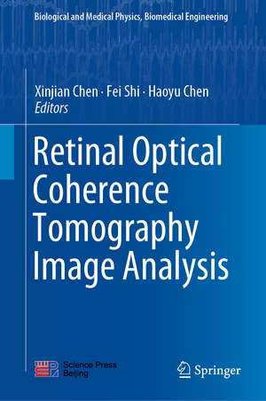
Retinal Optical Coherence Tomography Image Analysis: Biological and Medical Physics, Biomedical Engineering
Editat de Xinjian Chen, Fei Shi, Haoyu Chenen Limba Engleză Hardback – 16 iul 2019
Din seria Biological and Medical Physics, Biomedical Engineering
- 5%
 Preț: 1110.32 lei
 - 18%
 Preț: 1006.55 lei
 - 18%
 Preț: 960.78 lei
 - 18%
 Preț: 704.11 lei
 - 18%
 Preț: 967.40 lei
 - 18%
 Preț: 948.92 lei
 - 15%
 Preț: 641.71 lei
 - 15%
 Preț: 644.95 lei
 - 15%
 Preț: 665.08 lei
 - 18%
 Preț: 1669.16 lei
 - 18%
 Preț: 941.05 lei
 - 18%
 Preț: 956.81 lei
 - 18%
 Preț: 950.21 lei
 - 15%
 Preț: 636.80 lei
 - 18%
 Preț: 947.50 lei
 - 15%
 Preț: 636.80 lei
 -
 Preț: 397.01 lei
 - 18%
 Preț: 1236.99 lei
 - 15%
 Preț: 644.49 lei
 - 18%
 Preț: 946.55 lei
 - 15%
 Preț: 712.22 lei
 - 18%
 Preț: 952.89 lei
 - 18%
 Preț: 944.36 lei
 - 18%
 Preț: 1228.29 lei
 - 5%
 Preț: 1422.67 lei
 - 18%
 Preț: 1393.27 lei
 - 15%
 Preț: 651.19 lei
 - 18%
 Preț: 953.65 lei
 - 18%
 Preț: 955.88 lei
 - 15%
 Preț: 644.95 lei
 - 5%
 Preț: 1098.48 lei
 - 18%
 Preț: 959.19 lei
 - 15%
 Preț: 643.65 lei
 - 5%
 Preț: 1159.16 lei
 - 5%
 Preț: 1102.67 lei
 - 18%
 Preț: 952.09 lei
 - 18%
 Preț: 946.55 lei
 - 18%
 Preț: 952.09 lei
 - 15%
 Preț: 703.20 lei
 - 18%
 Preț: 953.65 lei
 - 5%
 Preț: 1008.45 lei
 - 15%
 Preț: 644.82 lei
 - 18%
 Preț: 956.03 lei
 - 15%
 Preț: 647.40 lei
Preț: 651.84 lei
Preț vechi: 766.87 lei
-15% Nou
Puncte Express: 978
Preț estimativ în valută:
124.77€ • 135.57$ • 104.87£
124.77€ • 135.57$ • 104.87£
Carte tipărită la comandă
Livrare economică 21 aprilie-05 mai
Preluare comenzi: 021 569.72.76
Specificații
ISBN-13: 9789811318245
ISBN-10: 9811318247
Pagini: 280
Ilustrații: XI, 385 p. 189 illus., 156 illus. in color.
Dimensiuni: 155 x 235 x 29 mm
Greutate: 0.74 kg
Ediția:1st ed. 2019
Editura: Springer Nature Singapore
Colecția Springer
Seria Biological and Medical Physics, Biomedical Engineering
Locul publicării:Singapore, Singapore
Cuprins
Clinical applications of retinal optical coherence tomography.- Fundamentals of optical coherence tomography.- Speckle noise reduction and enhancement for OCT images.- Reconstruction of retinal OCT images with sparse representation.- Segmentation of OCT scans using probabilistic graphical models.- Diagnostic capability of optical coherence tomography based quantitative analysis for various eye diseases and additional factors affecting morphological measurements.- Quantitative analysis of retinal layers' optical intensities based on optical coherence tomography.- Segmentation of optic disc and cup-to-disc ratio quantification based on OCT scans.- Choroidal OCT analytics.- Layer segmentation and analysis for retina with diseases.- Segmentation and visualization of drusen and geographic atrophy in SD-OCT images.- Segmentation of symptomatic exudate-associated derangements in 3D OCT images.- Modeling and prediction of chroidal neovascularization growth based on longitudinal OCT scans.
Notă biografică
Xinjian Chen, IEEE Senior Member, received his Ph.D. degree from the Institute of Automation, Chinese Academy of Sciences in 2006. After graduation, he worked on research projects with several prestigious groups: Microsoft Research Asia, Beijing, China (2006-2007); Medical Image Processing Group, University of Pennsylvania (2008-2009); Department of Radiology and Image Sciences, National Institutes of Health (2009-2011); and Department of Electrical and Computer Engineering, University of Iowa (2011-2012). In 2012, he joined the School of Electrical and Information Engineering, Soochow University where he serves as a Distinguished Professor and Director of Medical Image Processing, Analysis and Visualization Laboratory. Under his leadership, the laboratory has evolved with a strong group of 8 faculty members and 30 postgraduate students working towards PhD and MS programs. The laboratory has received more than 10 National and Provincial grants, including the prestigious Young Scientist grant with Xinjian as the PI from National Basic Research Program of China (973).
Dr. Chen is a recipient of the National Science Fund for Outstanding Young Scholars, China (2016), National One Thousand Young Talents Award, China (2012), Jiangsu Provincial High Level Creative Talents Award (2013), Jiangsu Provincial Peak Talents of Six Categories Award (2012), Beijing Science and Technology Advancement Award (2011), Chinese Academy of Sciences President Excellence Award (2006), Important Technology Innovation Award of China (2005), and National Technology Advancement Award of China (2004). His research interests include medical image processing and analysis, pattern recognition, machine learning, and their applications.
Dr. Chen has published more than 100 peer-reviewed papers in prestigious international journals and conferences including IEEE Transactions on Medical Imaging, IEEE Transactions on Biomedical Engineering, Medical Physics, IEEE Transactions Information Technology in Biomedicine, and Radiology, etc. He has served as Workshop Chair for MICCAI OMIA2014, Co-Chair for MICCAI OMIA2015, 2016, and 2017. He has also served as the Workshop Chair for International Medical Imaging Workshop at Suzhou 2013, 2014, 2015 and 2017. He has served as Associate Editor for IEEE Transactions on Medical Imaging, and IEEE Journal of Translational Engineering in Health and Medicine. He also holds 10 granted patents and 20 pending status patents.Fei Shi received her bachelor’s degree in Information and Electronics Engineering from Zhejiang University and her Ph.D. in Electrical Engineering from the Polytechnic University, Brooklyn, New York in 2006. She is currently an assistant professor at School of Electronics and Information Engineering, Soochow University. Her research interests include medical image processing, pattern recognition and time–frequency analysis. She has published more than 40 papers in international journals and conferences.
Haoyou Chen received an M.B.B.S. from Sun Yat-sen University in 2002, and M.D. from Zhongshan Ophthalmic Center, Sun Yat-sen University in 2008. He is currently a Consultant and Professor at the Joint Shantou International Eye Center, Shantou University and the Chinese University of Hong Kong. His research interests include ocular imaging and eye genetics. He has published 83 articles in international peer-reviewed journals and contributed to 6 book chapters. Dr. Chen serves as an Associate Editor for BMC Ophthalmology, editorial board member for Clinical Ophthalmology, and on the program committee for the 8th, 9th, 10th, 11th International Symposium of Ophthalmology (Hong Kong), and the 1st and 2nd MICCAI Workshop on Ophthalmic Medical Image Analysis. He has received several academic awards including Asia-Pacific Academy of Ophthalmology Achievement Award, Publons Peer Review Award, Youth Investigator Award in the 13th International Symposium on Retinal degeneration and a travel grant to Asia-ARVO 2015.
Textul de pe ultima copertă
This book introduces the latest optical coherence tomography (OCT) imaging and computerized automatic image analysis techniques, and their applications in the diagnosis and treatment of retinal diseases. Discussing the basic principles and the clinical applications of OCT imaging, OCT image preprocessing, as well as the automatic detection and quantitative analysis of retinal anatomy and pathology, it includes a wealth of clinical OCT images, and state-of-the-art research that applies novel image processing, pattern recognition and machine learning methods to real clinical data. It is a valuable resource for researchers in both medical image processing and ophthalmic imaging.
Caracteristici
Focuses on computerized automatic analysis of clinical optical coherence tomography (OCT) images Includes a wealth of examples of clinical OCT images for different retinal pathologies Offers comprehensive and in-depth descriptions of state-of-the-art image analysis methods applied to OCT images
