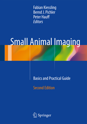
Small Animal Imaging: Basics and Practical Guide
Editat de Fabian Kiessling, Bernd J. Pichler, Peter Hauffen Limba Engleză Hardback – 7 iun 2017
| Toate formatele și edițiile | Preț | Express |
|---|---|---|
| Paperback (1) | 1128.39 lei 38-44 zile | |
| Springer International Publishing – 28 iul 2018 | 1128.39 lei 38-44 zile | |
| Hardback (1) | 1565.99 lei 38-44 zile | |
| Springer International Publishing – 7 iun 2017 | 1565.99 lei 38-44 zile |
Preț: 1565.99 lei
Preț vechi: 1648.42 lei
-5% Nou
Puncte Express: 2349
Preț estimativ în valută:
299.75€ • 325.70$ • 251.95£
299.75€ • 325.70$ • 251.95£
Carte tipărită la comandă
Livrare economică 16-22 aprilie
Preluare comenzi: 021 569.72.76
Specificații
ISBN-13: 9783319422008
ISBN-10: 3319422006
Pagini: 938
Ilustrații: XIX, 875 p. 396 illus., 284 illus. in color.
Dimensiuni: 178 x 254 mm
Greutate: 2.03 kg
Ediția:2nd ed. 2017
Editura: Springer International Publishing
Colecția Springer
Locul publicării:Cham, Switzerland
ISBN-10: 3319422006
Pagini: 938
Ilustrații: XIX, 875 p. 396 illus., 284 illus. in color.
Dimensiuni: 178 x 254 mm
Greutate: 2.03 kg
Ediția:2nd ed. 2017
Editura: Springer International Publishing
Colecția Springer
Locul publicării:Cham, Switzerland
Cuprins
Part I Role of small animal imaging: Non invasive imaging for supporting basic research.- Non invasive imaging in the pharmaceutical industry.- How to set up a small animal imaging unit.- Significance of non invasive imaging for animal protection.- Animal Models: From zebrafish to non human primates.
Part II Study planning and animal preparation: Institutional preconditions for small animal imaging.- Statistical considerations for animal imaging studies.- Animal anesthesia and monitoring.- Drug administration.
Part III Imaging modalities and probes: How to choose the right imaging modality.- How to identify suitable Biomarkers.- Concepts in diagnostic probe design.- X-ray and X-ray-CT.- CT contrast agents.- MR contrast agents.- MRS.- Hyperpolarized MRI.- Ultrasound.- Ultrasound contrast agents.- PET and SPECT.- Radiotracer I: Standard tracer.- Radiotracer II: Peptide-based radiopharmaceuticals.- Optical imaging.- Photoacoustic imaging.- Optical probes.- Multimodal imaging and image fusion.
Part IV Ex vivo validation methods:Methods for correlation between in vivo small ani-mal images and post mortem data: example brain.- In Vitro Methods for In Vivo Quantitation of PET and SPECT Imaging Probes: Autoradiography and Gamma Counting.
Part V: Data postprocessing: Qualitative and quantitative data analysis.- Guidelines for nuclear image analysis.- Kinetic modeling.- Data documentation systems.
Part VI: Special applications:Imaging in drug safety Evaluation.- Cell tracking and transplant imaging.- Imaging of metabolic diseases.- Imaging in developmental biology.- Imaging in gynecology research.- Imaging in cardiovascular research.- Imaging in neurology research I: Neuoroncology.- Imaging in neurology research II: Plasticity and cognitive networks.- Imaging in neurology research III: Neurodegenerative diseases.- Neurooptogenetics.- Imaging in oncology research.- Imaging in immunology research.- Imaging of infectious diseases.
Recenzii
“This book gives a detailed overview of the current situation regarding imaging of small animals with lots of tips and considerations and I can wholeheartedly recommend it.” (Maurits Jansen, RAD Magazine , May, 2018)
Notă biografică
Since 2008 Professor Dr. Fabian Kiessling is leading the Institute for Experimental Molecular Imaging at the Helmholtz Center of Applied Engineering of the RWTH-University in Aachen. Aim of his research is the development of novel diagnostic probes and imaging tools for a disease specific diagnosis and therapy monitoring. In this context, the main focus is on the investigation of angiogenesis related processes. Fabian Kiessling studied Medicine and graduated at the University of Heidelberg. Until the end of 2002, he worked as resident in the Department of Radiology at the German Cancer Research Center (DKFZ) in Heidelberg. In 2003 he changed to the Department of Medical Physics in Radiology of the DKFZ as leader of the Molecular Imaging group. In parallel he did his clinical training at different Departments of the University of Heidelberg and received the board certification as clinical Radiologist in 2007. Fabian Kiessling habilitated in experimental radiology in 2006. He is author of more than 240 publications and book chapters, received several research awards, among those the „Emil Salzer Price for Cancer Research” and the “Richtzenhain Price”. Shortly after his change to the RWTH-Aachen University he founded the invivoContrast GmbH together with Professor Matthias Bräutigam, which is distributing diagnostic probes for the preclinical market.
Textul de pe ultima copertă
This textbook is a practical guide to the use of small animal imaging in preclinical research that will assist in the choice of imaging modality and contrast agent and in study design, experimental setup, and data evaluation. All established imaging modalities are discussed in detail, with the assistance of numerous informative illustrations. While the focus of the new edition remains on practical basics, it has been updated to encompass a variety of emerging imaging modalities, methods, and applications. Additional useful hints are also supplied on the installation of a small animal unit, study planning, animal handling, and cost-effective performance of small animal imaging. Cross-calibration methods and data postprocessing are considered in depth. This new edition of Small Animal Imaging will be an invaluable aid for researchers, students, and technicians involved in research into and applications of small animal imaging.
Caracteristici
Provides invaluable practical guidance on the use of small animal imaging in preclinical research Offers detailed coverage of the available imaging modalities, including newly emerging ones, with the aid of numerous illustrations Will assist in the installation of a small animal unit, study planning, animal handling, and cost-effective imaging
