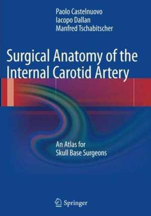
Surgical Anatomy of the Internal Carotid Artery: An Atlas for Skull Base Surgeons
Autor Paolo Castelnuovo, Iacopo Dallan, Manfred Tschabitscheren Limba Engleză Paperback – oct 2016
| Toate formatele și edițiile | Preț | Express |
|---|---|---|
| Paperback (1) | 765.09 lei 38-44 zile | |
| Springer Berlin, Heidelberg – oct 2016 | 765.09 lei 38-44 zile | |
| Hardback (1) | 726.32 lei 6-8 săpt. | |
| Springer Berlin, Heidelberg – 17 iun 2013 | 726.32 lei 6-8 săpt. |
Preț: 765.09 lei
Preț vechi: 805.36 lei
-5% Nou
Puncte Express: 1148
Preț estimativ în valută:
146.42€ • 151.70$ • 122.20£
146.42€ • 151.70$ • 122.20£
Carte tipărită la comandă
Livrare economică 18-24 martie
Preluare comenzi: 021 569.72.76
Specificații
ISBN-13: 9783662508398
ISBN-10: 3662508397
Pagini: 177
Ilustrații: XV, 162 p. 174 illus. in color.
Dimensiuni: 178 x 254 x 16 mm
Ediția:Softcover reprint of the original 1st ed. 2013
Editura: Springer Berlin, Heidelberg
Colecția Springer
Locul publicării:Berlin, Heidelberg, Germany
ISBN-10: 3662508397
Pagini: 177
Ilustrații: XV, 162 p. 174 illus. in color.
Dimensiuni: 178 x 254 x 16 mm
Ediția:Softcover reprint of the original 1st ed. 2013
Editura: Springer Berlin, Heidelberg
Colecția Springer
Locul publicării:Berlin, Heidelberg, Germany
Cuprins
Introduction.- Introduction of Internal Carotid Artery Classifications.- Cervical Segment.- Skull Base Segment.- Petrous.- Intracranial Segment.- Cavernous.- Supracavernous.
Recenzii
From the book reviews:
“This is a comprehensive atlas of the internal carotid artery, both intraoperatively and postmortem, outlining the branches of the ICA in full color. … This is best suited for neurosurgeons, fellows, and graduate residents in neurosurgery.” (Joseph J. Grenier, Amazon.com, March, 2015)
“This atlas provides unparalleled illustrations of all segments of the internal carotid artery from a skull base surgeon’s perspective. … It is most suitable for fellows and attendings in skull base surgery, especially those with experience in endoscopic and microscopic skull base surgery. … Rhinologists would find it valuable as they tend to avoid the internal carotid artery during training as otolaryngologists. … This is the only book solely dedicated to this topic that I have encountered.” (Opeyemi Daramola, Doody’s Book Reviews, October, 2013)
Textul de pe ultima copertă
This atlas provides all the basic and advanced information required by surgeons in order to understand fully the skull base anatomy. It is organized according to anatomo-surgical pathways to the hidden areas of the skull base. These pathways are described in step-by-step fashion with the aid of a wealth of color images and illustrations. The emphasis is on endoscopic anatomy, but in order to provide a holistic perspective, informative three-dimensional reconstructions are presented alongside the endoscopic images and radiologic images are included when appropriate. In effect, windows are opened on the anatomy so that the reader is guided on a journey throughout the skull base region. This anatomically oriented atlas will serve as an ideal learning tool for novice surgeons and will also prove an invaluable reference for the more experienced surgeon.
Caracteristici
Logical, step-by-step documentation of skull base anatomy with a wealth of color images and illustrations Emphasis on endoscopic anatomy and images Informative three-dimensional reconstructions Anatomic-radiologic correlations


