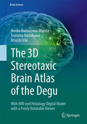
The 3D Stereotaxic Brain Atlas of the Degu: With MRI and Histology Digital Model with a Freely Rotatable Viewer: Brain Science
Autor Noriko Kumazawa-Manita, Tsutomu Hashikawa, Atsushi Irikien Limba Engleză Hardback – 20 oct 2018
| Toate formatele și edițiile | Preț | Express |
|---|---|---|
| Paperback (1) | 553.30 lei 38-44 zile | |
| Springer – 14 feb 2019 | 553.30 lei 38-44 zile | |
| Hardback (1) | 644.95 lei 3-5 săpt. | |
| Springer – 20 oct 2018 | 644.95 lei 3-5 săpt. |
Preț: 644.95 lei
Preț vechi: 758.77 lei
-15% Nou
Puncte Express: 967
Preț estimativ în valută:
123.41€ • 128.86$ • 101.91£
123.41€ • 128.86$ • 101.91£
Carte disponibilă
Livrare economică 25 martie-08 aprilie
Preluare comenzi: 021 569.72.76
Specificații
ISBN-13: 9784431566137
ISBN-10: 4431566139
Pagini: 120
Ilustrații: IX, 144 p. 198 illus., 144 illus. in color.
Dimensiuni: 178 x 254 mm
Greutate: 0.54 kg
Ediția:1st ed. 2018
Editura: Springer
Colecția Springer
Seria Brain Science
Locul publicării:Tokyo, Japan
ISBN-10: 4431566139
Pagini: 120
Ilustrații: IX, 144 p. 198 illus., 144 illus. in color.
Dimensiuni: 178 x 254 mm
Greutate: 0.54 kg
Ediția:1st ed. 2018
Editura: Springer
Colecția Springer
Seria Brain Science
Locul publicării:Tokyo, Japan
Cuprins
Chapter 1: Introduction, Materials and Methods, and References.- Chapter 2: List of Structures.- Chapter 3: The Degu Brain Atlas.- Chapter 4: SG-eye Operation Manual.- Index of Structures and Abbreviations.
Notă biografică
Noriko Kumazawa-Manita
Laboratory for Symbolic Cognitive Development, RIKEN Brain Science Institute, Japan
Tsutomu Hashikawa
Laboratory for Symbolic Cognitive Development, RIKEN Brain Science Institute, Japan
Atsushi Iriki
Laboratory for Symbolic Cognitive Development, RIKEN Brain Science Institute, Japan
Laboratory for Symbolic Cognitive Development, RIKEN Brain Science Institute, Japan
Tsutomu Hashikawa
Laboratory for Symbolic Cognitive Development, RIKEN Brain Science Institute, Japan
Atsushi Iriki
Laboratory for Symbolic Cognitive Development, RIKEN Brain Science Institute, Japan
Textul de pe ultima copertă
This is the first digital atlas of the degu brain with microscopic features simultaneously in Nissl sections and magnetic resonance imaging (MRI). As an experimental animal model, the degu contributes to a variety of medical research fields in diabetes, hyperglycemia, pancreatic function, and adaptation to high altitude, among others. Recently the degu has gained increasing importance in the field of neuroscience, particularly in studies evaluating the relationship between sociality and cognitive brain functions, and in studies pertaining to the evolutional aspects of the acquisition of tool-use abilities. Furthermore, aging-related brain dysfunction in humans can be studied using this animal model in addition to mammals with much longer lifespans. This brain atlas is constructed to provide histological and volume-rendered information simultaneously, fitting with any spatial coordination in brain positioning. It can be a useful guide to degus as well as to other rodents for studies ofbrain structures conducted using MRI or other contemporary examination methods with volume-rendering functions.
Caracteristici
Helps the reader to identify individual brain structures in the degu by including numerous histological images with delineation lines Presents anatomical information simultaneously from Nissl and magnetic resonance images Readers can enjoy 3D views of the reconstructed brain and freely oriented planes of sections at a dedicated website described in the book


