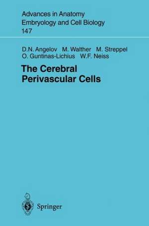
The Cerebral Perivascular Cells: Advances in Anatomy, Embryology and Cell Biology, cartea 147
Autor Doychin N. Angelov, Michael Walther, Michael Streppel, Orlando Guntinas-Lichius, Wolfram F. Neissen Limba Engleză Paperback – 21 sep 1998
Din seria Advances in Anatomy, Embryology and Cell Biology
- 5%
 Preț: 1146.33 lei
 - 5%
 Preț: 721.19 lei
 - 15%
 Preț: 637.13 lei
 -
 Preț: 381.81 lei
 - 15%
 Preț: 644.95 lei
 - 5%
 Preț: 1025.16 lei
 - 15%
 Preț: 689.97 lei
 - 15%
 Preț: 577.07 lei
 - 15%
 Preț: 580.36 lei
 - 5%
 Preț: 393.51 lei
 -
 Preț: 408.66 lei
 -
![Die Schlüpfdrüse der Geburtshelferkröte (Alytes o. obstetricans [LAURENTI]) und anderer Froschlurche](https://i4.books-express.ro/bs/9783662239742/die-schluepfdruese-der-geburtshelferkroete-alytes-o-obstetricans-laurenti-und-anderer-froschlurche.jpg) Preț: 408.27 lei
Preț: 408.27 lei - 5%
 Preț: 1090.61 lei
 - 5%
 Preț: 705.11 lei
 - 5%
 Preț: 706.04 lei
 - 5%
 Preț: 357.61 lei
 - 5%
 Preț: 704.59 lei
 - 5%
 Preț: 705.11 lei
 - 5%
 Preț: 359.42 lei
 - 5%
 Preț: 711.52 lei
 - 15%
 Preț: 635.47 lei
 - 15%
 Preț: 631.72 lei
 - 15%
 Preț: 633.35 lei
 - 15%
 Preț: 632.37 lei
 - 5%
 Preț: 706.60 lei
 - 15%
 Preț: 631.07 lei
 - 5%
 Preț: 707.13 lei
 - 5%
 Preț: 707.33 lei
 - 5%
 Preț: 359.60 lei
 - 5%
 Preț: 707.69 lei
 - 5%
 Preț: 707.13 lei
 - 5%
 Preț: 708.06 lei
 - 5%
 Preț: 706.41 lei
 - 5%
 Preț: 708.78 lei
 - 5%
 Preț: 705.68 lei
 - 5%
 Preț: 705.11 lei
 - 5%
 Preț: 706.77 lei
 - 15%
 Preț: 635.15 lei
 - 15%
 Preț: 631.07 lei
 - 5%
 Preț: 706.77 lei
 - 5%
 Preț: 706.04 lei
 - 5%
 Preț: 710.79 lei
 - 5%
 Preț: 705.32 lei
 - 15%
 Preț: 633.19 lei
 - 15%
 Preț: 629.09 lei
 - 15%
 Preț: 633.53 lei
 - 15%
 Preț: 632.70 lei
 - 15%
 Preț: 633.68 lei
 - 18%
 Preț: 773.72 lei
 - 15%
 Preț: 630.43 lei
Preț: 631.07 lei
Preț vechi: 742.43 lei
-15% Nou
Puncte Express: 947
Preț estimativ în valută:
120.76€ • 129.13$ • 100.68£
120.76€ • 129.13$ • 100.68£
Carte tipărită la comandă
Livrare economică 17 aprilie-01 mai
Preluare comenzi: 021 569.72.76
Specificații
ISBN-13: 9783540646389
ISBN-10: 3540646388
Pagini: 104
Ilustrații: XI, 90 p. 23 illus.
Dimensiuni: 155 x 235 x 5 mm
Greutate: 0.16 kg
Ediția:Softcover reprint of the original 1st ed. 1998
Editura: Springer Berlin, Heidelberg
Colecția Springer
Seria Advances in Anatomy, Embryology and Cell Biology
Locul publicării:Berlin, Heidelberg, Germany
Public țintă
ResearchCuprins
1 Introduction.- 1.1 Antigen Presentation and Antigen Presenting Cells.- 1.2 Antigen Presentation within the CNS.- 1.3 Microglia Might Be the Cerebral Antigen Presenting Cells.- 1.4 Theories on the Antigen Presentation Site.- 1.5 Questions Still Open.- 1.6 Methodological Approach.- 2 Materials and Methods.- 2.1 Animals.- 2.2 Overview of Animal Experiments.- 2.3 Surgery.- 2.4 Number of Sprouting Neurons After Retrograde Tracing with HRP.- 2.5 Number of Surviving Neurons After Resection of the Facial Nerve.- 2.6 Neuronophagic Microglia Identified by Vital Labeling with Fluoro-Gold.- 3 Results.- 3.1 Resection of 10 mm of the Facial Nerve Causes a Slow Loss of Facial Motoneurons in the Adult Rat.- 3.2 No Breakdown of the Blood — Brain Barrier or Passage of Unprimed Lymphocytes into Brain Tissue After Facial Nerve Resection.- 3.3 Fluoro-Gold Labeling of Motoneurons, Phagocytic Microglia and Perivascular Cells.- 3.4 Time Course of Existence and Migration of Fluoro-Gold-Labeled Neuronophages.- 3.5 Immunocytochemistry of the Fluoro-Gold-Labeled Neuronophages.- 4 Discussion.- 4.1 Oligodendrocytes and Astrocytes Are Not the APC of the Brain.- 4.2 Microglia Are Not the APC of the Brain.- 4.3 The Perivascular Cells Are the APC of the Brain.- 5 Summary.- References.
Textul de pe ultima copertă
Following an exhaustive literature review on the global issue of intracerebral presentation of antigen, this monograph summarizes results from voluminous work to establish which indigenous cerebral cells might present (auto)antigen to the immune system and thus initiate an (auto)immune reaction. Employing the combination of (a) a lesion model in which neuronal degeneration and neuronophagia are caused without disruption of the blood--brain barrier, (b) stable labeling of the neuronophages via phagocytosis of the permanent nontoxic fluorescent marker Fluoro-Gold from preloaded neurons, and (c) immunocytochemical identification of all FG-labeled brain neuronophages, the authors provide evidence that the only cells in the rat CNS which can be regarded as the resident antigen presenting cells of the brain are perivascular cells.
