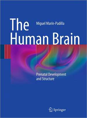
The Human Brain: Prenatal Development and Structure
Autor Miguel Marín-Padillaen Limba Engleză Hardback – 10 noi 2010
| Toate formatele și edițiile | Preț | Express |
|---|---|---|
| Paperback (1) | 701.90 lei 38-44 zile | |
| Springer Berlin, Heidelberg – 23 aug 2016 | 701.90 lei 38-44 zile | |
| Hardback (1) | 970.18 lei 38-44 zile | |
| Springer Berlin, Heidelberg – 10 noi 2010 | 970.18 lei 38-44 zile |
Preț: 970.18 lei
Preț vechi: 1021.24 lei
-5% Nou
Puncte Express: 1455
Preț estimativ în valută:
185.64€ • 194.35$ • 153.61£
185.64€ • 194.35$ • 153.61£
Carte tipărită la comandă
Livrare economică 01-07 aprilie
Preluare comenzi: 021 569.72.76
Specificații
ISBN-13: 9783642147234
ISBN-10: 3642147232
Pagini: 100
Ilustrații: XII, 145 p.
Dimensiuni: 193 x 260 x 13 mm
Greutate: 0.61 kg
Ediția:2011
Editura: Springer Berlin, Heidelberg
Colecția Springer
Locul publicării:Berlin, Heidelberg, Germany
ISBN-10: 3642147232
Pagini: 100
Ilustrații: XII, 145 p.
Dimensiuni: 193 x 260 x 13 mm
Greutate: 0.61 kg
Ediția:2011
Editura: Springer Berlin, Heidelberg
Colecția Springer
Locul publicării:Berlin, Heidelberg, Germany
Public țintă
ResearchCuprins
Mammalian Cerebral Cortex: Embryonic Devolpment and Cytoarchitecture.-Human Motor Cortex: Development and Cytoarchitecture.-The Mammalian Pyramidal Neuron: Development, Structure and Function.-Human Motor Cortex First Lamina: Devolpment and Cytoarchitecture.-Human Motor Cortex Excitatory/ Inhibitory Neuronal Systems: Devolpment and Cytoarchitecture.-Human Cerebral Cortex Intrinsic Microvascular System: Devolpment and Cytoarchitecture.-Human Motor Cortex First Lamina and Gray Matter Special Astrocytes: Devolpment and Cytoarchitecture.-New Developmental Cytoarchitectonics Therory and Nomenclature.- Epilogue.- Cat Motor Cortex: Devolpment and Cytoarchitecture.-Appendix: The Rapid Golgi Reaction - A Personal Quest.
Recenzii
From the reviews:
“Marín-Padilla’s recent monograph will be of great significance for anyone now embarking on the study of cortical development; it is an obligatory source for anyone working in the field of cortical development with an interest in clinical translation. … His presentations were colorful and elegant, using forceful, captivating and entertaining rhetoric. … great value for future generations of paediatric neuropathologists and researchers … . I enjoyed reading the book … . I envisage keeping this book on a shelf in my laboratory … .” (Zoltán Molnár, Brain, Vol. 134, 2011)
“In 143 beautifully written and illustrated pages, the author presents the organization and development of the human and feline motor cortex, as well as personal reflections on his scientific quest with the Golgi stain. … This book is relevant to neuropathologists because it presents from the 3-dimensional perspective the normative organization of the developing human cortex as a basis for interpretation of the multiple destructive and malformative disorders of the embryo and fetus. The book is relevant to clinicians … . ” (Hannah C. Kinney, Journal of Neuropathology and Experimental Neurology, Vol. 71 (2), February, 2012)
Notă biografică
Dr. Miguel Marín-Padilla is Professor Emeritus of Pathology and of Pediatrics at Dartmouth Medical School (Hanover, New Hampshire, USA). He obtained his Medical Degree in Spain (Granada University, 1955), practice Pediatrics for one year but interested in pediatric research immigrate to USA (1956). In USA, he completed Clinical Internship and several years of Pathology Residencies (Mallory Institute of Pathology, Boston), with emphasis in both Developmental and Pediatric Pathology (1957-1961) and was Teaching Fellow in Pathology in both Boston and Harvard Medical Schools (1960-62). He pursued an Academic Career, from Instructor to full Professor in both Pathology and Pediatrics at Dartmouth Medical School (1962-2000). He did a Special Neurohistology Fellowships at the Cajal Institute (Madrid) to study Cajal old Golgi preparations and learn the method (1966-67) and was Visiting Neuroscientist at University of Alcalá de Henares (Spain, 1994) and at the Mayo Clinic (1999). His research efforts has resulted in 180 publications, including abstracts, peer review papers, collaborative works, chapters in books and books. His developmental studies are recognized the world over and he has received various honors for his teaching and for his research efforts, the most notable are:Alpha Omega Alpha (USA Honor Medical Society, 1981); Best teacher award (1988,1996) and Faculty Speaker at Commencements (1985, 1991) at Dartmouth Medical School; Cajal Medal (USA Cajal Club, 1990); Gold Medal ‘Aureliano Maestre de San Juan’ of the University of Granada (1997), Honor Member of the Spanish Neurology (1987) and Pediatric Neurology Societies (2000), The Jacod Javist Neuroscience Investigator Award (USA, 1989), Honor Member of the Royal Academy of Medicine (Murcia, 2001), the Gold Medal of the Community of Murcia (2001) and has been a Candidate to the Principe de Asturias in Sciences (2002), “The Miguel Marín-Padilla Award, Excellence in Pathology”. Annual Lectureship andAward. Dartmouth Medical School (2006). His research has been supported by Grants of the U.S. National Institute of Health from 1962 to 1999.
Textul de pe ultima copertă
This book is unique among the current literature in that it systematically documents the prenatal structural development of the human brain. It is based on lifelong study using essentially a single staining procedure, the classic rapid Golgi procedure, which ensures an unusual and desirable uniformity in the observations. The book is amply illustrated with 81 large, high-quality color photomicrographs never previously reproduced. These photomicrographs, obtained at 6, 7, 11, 15, 18, 20, 25, 30, 35, and 40 weeks of gestation, offer a fascinating insight into the sequential prenatal development of neurons, blood vessels, and glia in the human brain.
Caracteristici
Systematically documents the prenatal structural development of neurons, blood vessels, and glia in the human brain Based on lifelong study using essentially a single staining procedure, the classic rapid Golgi procedure Amply illustrated with 81 large, high-quality color photomicrographs obtained from 6 to 40 weeks of gestation and never previously reproduced Includes supplementary material: sn.pub/extras
