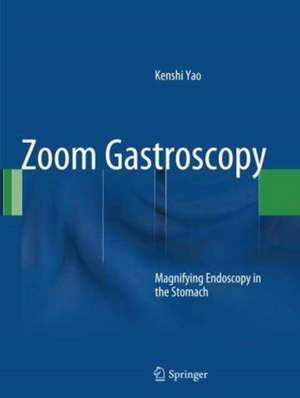
Zoom Gastroscopy: Magnifying Endoscopy in the Stomach
Autor Kenshi Yaoen Limba Engleză Paperback – 17 sep 2016
| Toate formatele și edițiile | Preț | Express |
|---|---|---|
| Paperback (1) | 1184.05 lei 38-44 zile | |
| Springer – 17 sep 2016 | 1184.05 lei 38-44 zile | |
| Hardback (1) | 1472.17 lei 3-5 săpt. | |
| Springer – 11 dec 2013 | 1472.17 lei 3-5 săpt. |
Preț: 1184.05 lei
Preț vechi: 1246.37 lei
-5% Nou
Puncte Express: 1776
Preț estimativ în valută:
226.61€ • 235.95$ • 191.51£
226.61€ • 235.95$ • 191.51£
Carte tipărită la comandă
Livrare economică 07-13 martie
Preluare comenzi: 021 569.72.76
Specificații
ISBN-13: 9784431561187
ISBN-10: 4431561188
Pagini: 230
Ilustrații: XV, 215 p. 382 illus., 168 illus. in color.
Dimensiuni: 210 x 279 x 18 mm
Greutate: 6.43 kg
Ediția:Softcover reprint of the original 1st ed. 2014
Editura: Springer
Colecția Springer
Locul publicării:Tokyo, Japan
ISBN-10: 4431561188
Pagini: 230
Ilustrații: XV, 215 p. 382 illus., 168 illus. in color.
Dimensiuni: 210 x 279 x 18 mm
Greutate: 6.43 kg
Ediția:Softcover reprint of the original 1st ed. 2014
Editura: Springer
Colecția Springer
Locul publicării:Tokyo, Japan
Cuprins
Chapter 1 Basic principles for the interpretation of magnified endoscopic (ME) findings: vessels (V) plus surface (S) classification system.- Chapter 2 Magnifying endoscopy (ME) of the stomach targeting the microvascular architecture.- Chapter 3 Magnifying ratio and resolution in electronic endoscopy systems.- Chapter 4 Magnified endoscopic (ME) findings of the normal gastric mucosa.- Chapter 5 Chronic gastritis– gastric body mucosal patterns identified using magnifying endoscopy (ME).- Chapter 6 Microvascular architecture characteristic of early gastric cancer as visualized by magnifying endoscopy (ME), and clinical applications.- Chapter 7 Principles of magnifying endoscopy with narrow-band imaging (M-NBI).- Chapter 8 Microanatomies as visualized using magnifying endoscopy with narrow band imaging (M-NBI) in the stomach.- Chapter 9 The proposed vessels plus surface (VS) classification system – principles for interpretation of magnifying endoscopy with narrow-band imaging (M-NBI) findings.- Chapter 10 Light blue crests (LBCs) and white opaque substance (WOS).- Chapter 11 Clinical applications of magnifying endoscopy with narrow band imaging (M-NBI) of the stomach.- Chapter 12 The vessels plus surface (VS) classification system in the diagnosis of early gastric cancer.- Chapter 13 Analysis and interpretation of magnifying endoscopy with narrow band imaging (M-NBI) findings in gastric epithelial tumors (early gastric cancer and adenoma) stratified for Paris classification of macroscopic appearance.- Chapter 14 New clinical applications and advantages for magnifying endoscopy (ME) with narrow band imaging (NBI) in the diagnosis of early gastric cancer –clinical usefulness, limitations and clinical strategies in preoperative determination of tumor margins, according to degree of difficultycategory.
Textul de pe ultima copertă
Since magnifying endoscopy of the stomach, or zoom gastroscopy, was first performed at the beginning of the 21st century , there have been numerous findings in the field worldwide. The author of this volume has developed a standardized magnifying endoscopy technique that enables endoscopists to constantly visualize the microanatomy (subepithelial capillaries and the epithelial structure) within the stomach. With this technology, it has become easy to obtain magnified endoscopic findings. Techniques differ, however, depending upon the endoscopist. Endoscopists around the world do not have a standardized magnifying endoscopy technique, which is mandatory for the scientific analysis of their findings. In addition, there is no logical explanation for how and which microanatomies are visualized by magnifying endoscopy with narrow-band imaging in the glandular epithelium of the stomach. This inconsistency results in considerable confusion among researchers and clinicians and a lack of terminology for the purposes of analysis. As magnifying endoscopy with narrow-band imaging (NBI) becomes more popular throughout the world, there is increased demand for a specialized reference book focused on this subject. The aims of the present volume, Zoom Gastroscopy: Magnifying Endoscopy in the Stomach, are manifold: (1) to illustrate the standard magnifying endoscopic technique that can visualize subepithelial mucosal microvessels as small as capillaries, the smallest unit of blood vessels in the human body, (2) to explain the optical phenomena and principles for NBI and its application to magnifying endoscopy, (3) to clarify which microanatomies are visualized by magnifying endoscopy with NBI and how this is achieved, and (4) to present a diagnostic system so called “VS classification system” for early gastric neoplasias. With a single author, the book consistently uses uniform anatomical terms to describe endoscopic findings, providing an essential reference forendoscopists in the world.
Caracteristici
Shows standard magnifying endoscopic technique in the stomach Explains the optical phenomena and principle for narrow-band imaging (NBI) and its application to magnifying endoscopy in the stomach Clarifies which microanatomies are visualized by magnifying endoscopy with NBI and how this is achieved in the glandular epithelium of the stomach Presents a diagnostic system so called “VS classification system” for early gastric neoplasias


