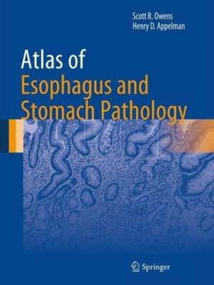
Atlas of Esophagus and Stomach Pathology: Atlas of Anatomic Pathology
Autor Scott R. Owens, Henry D. Appelmanen Limba Engleză Hardback – 7 sep 2013
Authored by nationally and internationally recognized pathologists, Atlas of Esophagus and Stomach Pathology is a valuable tool for both pathologists-in-training seeking to make “new acquaintances”, and practicing surgical pathologists in need of a quick visual reference in recalling “old friends” in the world of diagnostic gastrointestinal pathology.
| Toate formatele și edițiile | Preț | Express |
|---|---|---|
| Paperback (1) | 713.67 lei 38-44 zile | |
| Springer – 23 aug 2016 | 713.67 lei 38-44 zile | |
| Hardback (1) | 1110.32 lei 3-5 săpt. | |
| Springer – 7 sep 2013 | 1110.32 lei 3-5 săpt. |
Din seria Atlas of Anatomic Pathology
- 5%
 Preț: 1432.17 lei
 - 5%
 Preț: 1108.51 lei
 - 5%
 Preț: 1966.17 lei
 - 5%
 Preț: 1114.91 lei
 - 5%
 Preț: 1113.99 lei
 - 5%
 Preț: 739.13 lei
 - 5%
 Preț: 1040.68 lei
 - 5%
 Preț: 1442.42 lei
 - 5%
 Preț: 413.57 lei
 - 5%
 Preț: 1285.69 lei
 - 5%
 Preț: 1345.79 lei
 - 5%
 Preț: 1251.48 lei
 - 5%
 Preț: 1311.40 lei
 - 5%
 Preț: 1199.79 lei
 - 5%
 Preț: 849.44 lei
 - 5%
 Preț: 740.05 lei
Preț: 1110.32 lei
Preț vechi: 1168.76 lei
-5% Nou
Puncte Express: 1665
Preț estimativ în valută:
212.45€ • 222.42$ • 175.80£
212.45€ • 222.42$ • 175.80£
Carte disponibilă
Livrare economică 15-29 martie
Preluare comenzi: 021 569.72.76
Specificații
ISBN-13: 9781461480839
ISBN-10: 1461480833
Pagini: 195
Ilustrații: X, 195 p. 373 illus. in color.
Dimensiuni: 210 x 279 x 15 mm
Greutate: 0.82 kg
Ediția:2014
Editura: Springer
Colecția Springer
Seria Atlas of Anatomic Pathology
Locul publicării:New York, NY, United States
ISBN-10: 1461480833
Pagini: 195
Ilustrații: X, 195 p. 373 illus. in color.
Dimensiuni: 210 x 279 x 15 mm
Greutate: 0.82 kg
Ediția:2014
Editura: Springer
Colecția Springer
Seria Atlas of Anatomic Pathology
Locul publicării:New York, NY, United States
Public țintă
Professional/practitionerCuprins
Normal Histologic Anatomy and Common Variations.- General Microscopic Changes.- Noninfectious Inflammatory Conditions.- Infectious Esophagitis and Organisms.- Benign Squamous Proliferations.- Squamous Intraepithelial Neoplasia.- Invasive Squamous Cell Carcinoma.- Unusual Carcinomas, Including Squamous Variants.- Nonneoplastic Barrett's Mucosa.- Dysplasia and Dysplasia Mimics in Barrett's Mucosa.- Invasive Adenocarcinoma in Barrett's Mucosa.- Mesenchymal and Melanocytic Proliferations.- Esophageal Odds and Ends.- Gastric Cardiac Mucosa.- Normal Biopsy Anatomy/Histology and General Changes.- General Microscopic Changes and Findings to Ignore.- Structural Abnormalities/Heterotopias.- Vascular Abnormalities.- Toxic and Medication-Induced Conditions.- Noninfectious Inflammatory and Systemic Diseases Affecting the Stomach.- Infectious Diseases and Organisms.- Hyperplastic/Metaplastic Conditions.- Benign Tumors and Polyps.- Epithelial Dysplasia and Adenomas.- Carcinoma of the Stomach.- Carcinoid Tumor of the Stomach.- Mesenchymal Neoplasms.- Hematolymphoid Neoplasms.- Gastric Metastases.
Recenzii
From the reviews:
“This unique atlas emphasizes the real world experience of pathologists studying the upper gastrointestinal tract … . This atlas can be an invaluable tool for both pathologists-in-training and seasoned, practicing pathologists. … unique book that offers a more realistic approach to commonly encountered upper gastrointestinal biopsy specimens with less than perfect, real-world images and the personal experiences of internationally recognized gastrointestinal pathologists.” (Deepthi S. Rao, Doody’s Book Reviews, March, 2014)
“This unique atlas emphasizes the real world experience of pathologists studying the upper gastrointestinal tract … . This atlas can be an invaluable tool for both pathologists-in-training and seasoned, practicing pathologists. … unique book that offers a more realistic approach to commonly encountered upper gastrointestinal biopsy specimens with less than perfect, real-world images and the personal experiences of internationally recognized gastrointestinal pathologists.” (Deepthi S. Rao, Doody’s Book Reviews, March, 2014)
Notă biografică
Scott R. Owens, MD
University of Michigan, Department of Pathology, Ann Arbor, MI, USA
Henry D. Appelman, MD
University of Michigan, Department of Pathology, Ann Arbor, MI, USA
University of Michigan, Department of Pathology, Ann Arbor, MI, USA
Henry D. Appelman, MD
University of Michigan, Department of Pathology, Ann Arbor, MI, USA
Textul de pe ultima copertă
Atlas of Esophagus and Stomach Pathology provides an image-based resource for those studying normal histology of the upper gastrointestinal tract, as well as the microscopic manifestations of developmental abnormalities, toxic insults, infectious diseases, inflammatory and autoimmune conditions, and neoplasia in the esophagus and stomach. Because modern gastrointestinal pathology practice centers on specimens obtained during endoscopic examination, the atlas focuses on biopsy pathology, providing “real-world” microscopic images and ancillary diagnostic studies for most commonly-encountered abnormalities and diseases affecting these two organs. The book is supplemented with endoscopic and special study images.
Authored by nationally and internationally recognized pathologists, Atlas of Esophagus and Stomach Pathology is a valuable tool for both pathologists-in-training seeking to make “new acquaintances”, and practicing surgical pathologists in need of a quick visual reference in recalling “old friends” in the world of diagnostic gastrointestinal pathology.
Authored by nationally and internationally recognized pathologists, Atlas of Esophagus and Stomach Pathology is a valuable tool for both pathologists-in-training seeking to make “new acquaintances”, and practicing surgical pathologists in need of a quick visual reference in recalling “old friends” in the world of diagnostic gastrointestinal pathology.
Caracteristici
Authored by nationally and internationally recognized pathologists Features visual diagnostic criteria with detailed figure legends A quick reference guide for the diagnosis of usual and unusual diseases Supplemented with endoscopic and special study images Includes supplementary material: sn.pub/extras
