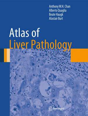
Atlas of Liver Pathology: Atlas of Anatomic Pathology
Autor Anthony W.H. Chan, Alberto Quaglia, Beate Haugk, Alastair Burten Limba Engleză Hardback – 7 noi 2013
Authored by nationally and internationally recognized pathologists, Atlas of Liver Pathology is a valuable resource that serves as a quick reference guide for the diagnosis of usual and unusual diseases.
| Toate formatele și edițiile | Preț | Express |
|---|---|---|
| Paperback (1) | 723.86 lei 38-44 zile | |
| Springer – oct 2016 | 723.86 lei 38-44 zile | |
| Hardback (1) | 1114.91 lei 3-5 săpt. | |
| Springer – 7 noi 2013 | 1114.91 lei 3-5 săpt. |
Din seria Atlas of Anatomic Pathology
- 5%
 Preț: 1432.17 lei
 - 5%
 Preț: 1108.51 lei
 - 5%
 Preț: 1966.17 lei
 - 5%
 Preț: 1110.32 lei
 - 5%
 Preț: 1113.99 lei
 - 5%
 Preț: 739.13 lei
 - 5%
 Preț: 1040.68 lei
 - 5%
 Preț: 1442.42 lei
 - 5%
 Preț: 413.58 lei
 - 5%
 Preț: 1285.73 lei
 - 5%
 Preț: 1345.82 lei
 - 5%
 Preț: 1251.52 lei
 - 5%
 Preț: 1311.42 lei
 - 5%
 Preț: 1199.81 lei
 - 5%
 Preț: 849.45 lei
 - 5%
 Preț: 740.05 lei
Preț: 1114.91 lei
Preț vechi: 1173.60 lei
-5% Nou
Puncte Express: 1672
Preț estimativ în valută:
213.39€ • 230.34$ • 178.93£
213.39€ • 230.34$ • 178.93£
Carte disponibilă
Livrare economică 29 martie-12 aprilie
Preluare comenzi: 021 569.72.76
Specificații
ISBN-13: 9781461491132
ISBN-10: 1461491134
Pagini: 231
Ilustrații: X, 248 p. 706 illus., 220 illus. in color.
Dimensiuni: 210 x 279 x 17 mm
Greutate: 0.93 kg
Ediția:2014
Editura: Springer
Colecția Springer
Seria Atlas of Anatomic Pathology
Locul publicării:New York, NY, United States
ISBN-10: 1461491134
Pagini: 231
Ilustrații: X, 248 p. 706 illus., 220 illus. in color.
Dimensiuni: 210 x 279 x 17 mm
Greutate: 0.93 kg
Ediția:2014
Editura: Springer
Colecția Springer
Seria Atlas of Anatomic Pathology
Locul publicării:New York, NY, United States
Public țintă
Professional/practitionerCuprins
Normal, Variants, and Methods.- General Processes.- Developmental Abnormality.- Metabolic Liver Disease.- Fatty Liver Disease.- Viral Liver Disease.- Nonviral Infectious Liver Disease.- Drug-Induced Liver Injury.- Autoimmune Hepatitis.- Biliary Disease.- Vascular Disorders.- Premalignant Lesion.- Neoplasm-like Liver Lesion.- Epithelial Liver Neoplasm.- Nonepithelial Liver Neoplasm.- Obstetric Liver Disease.- Transplantation Pathology.
Recenzii
From the reviews:
“This book focuses on pictures of normal liver histology and the most common liver diseases. … It is intended for use by students, residents, and general pathologists in the interpretation of liver biopsy histology. … The best part of the book is that it is mostly pictures with short descriptions that are detailed enough to be very helpful and provide a differential/mimickers of the entity shown. … The pictures are clear, numerous, and excellent examples of the liver histopathology.” (Hana Albrecht, Doody’s Book Reviews, May, 2014)
“This book focuses on pictures of normal liver histology and the most common liver diseases. … It is intended for use by students, residents, and general pathologists in the interpretation of liver biopsy histology. … The best part of the book is that it is mostly pictures with short descriptions that are detailed enough to be very helpful and provide a differential/mimickers of the entity shown. … The pictures are clear, numerous, and excellent examples of the liver histopathology.” (Hana Albrecht, Doody’s Book Reviews, May, 2014)
Notă biografică
Anthony W.H. Chan, BMedSc, MBChB, FRCPA, FHKCPath, FHKAM (Pathology)
Associate Consultant, The Chinese University of Hong Kong, Prince of Wales Hospital, Department of Anatomical and Cellular Pathology, Hong Kong, China
Alberto Quaglia, MD, PhD, FRCPath
Lead Consultant Histopathologist, King’s College Hospital, Institute of Liver Studies, London, UK
Beate Haugk, MD, FRCPath
Consultant Histopathologist, Royal Victoria Infirmary, Department of Cellular Pathology, Newcastle upon Tyne, UK
Alastair Burt, BSc(Hons), MBChB, MD(Hons), FRCPath, FRCP, FSB
The University of Adelaide, Head, School of Medicine, Dean of Medicine, Adelaide, Australia
Textul de pe ultima copertă
The liver is a complex organ due to its unique microscopic structure, intricate metabolic functions and susceptibility to a wide variety of insults, manifesting in countless histological patterns. Atlas of Liver Pathology considers both changes seen in medical liver biopsies as well as lesional biopsies when the specimen has been taken from a mass. The book starts by reviewing normal structure and its variants and the optimal approaches for the preparation of histological sections for diagnostic liver pathology. The following chapters are dedicated to developmental, metabolic, infectious, drug related, autoimmune, biliary, vascular and neoplastic disorders. Two sections on liver pathology in pregnancy and transplantation conclude the work. Macroscopic illustrations are included where appropriate. All photographs are complemented by legends describing the picture and providing relevant related information.
Authored by nationally and internationally recognized pathologists, Atlas of Liver Pathology is a valuable resource that serves as a quick reference guide for the diagnosis of usual and unusual diseases.
Authored by nationally and internationally recognized pathologists, Atlas of Liver Pathology is a valuable resource that serves as a quick reference guide for the diagnosis of usual and unusual diseases.
Caracteristici
Authored by nationally and internationally recognized pathologists Features visual diagnostic criteria with detailed figure legends A quick reference guide for the diagnosis of usual and unusual diseases Supplemented with radiographic and special study images
