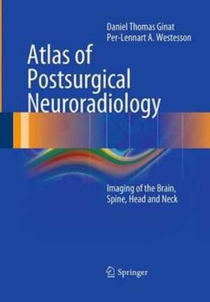
Atlas of Postsurgical Neuroradiology: Imaging of the Brain, Spine, Head, and Neck
Autor Daniel Thomas Ginat, Per-Lennart A. Westessonen Limba Engleză Paperback – 23 aug 2016
Toate formatele și edițiile
| Toate formatele și edițiile | Preț | Express |
|---|---|---|
| Paperback (1) | 1243.79 lei 6-8 săpt. | |
| Springer Berlin, Heidelberg – 23 aug 2016 | 1243.79 lei 6-8 săpt. | |
| Hardback (1) | 2275.34 lei 39-44 zile | |
| Springer International Publishing – 12 iul 2017 | 2275.34 lei 39-44 zile |
Preț: 1243.79 lei
Preț vechi: 1309.26 lei
-5% Nou
Puncte Express: 1866
Preț estimativ în valută:
238.02€ • 248.81$ • 201.13£
238.02€ • 248.81$ • 201.13£
Carte tipărită la comandă
Livrare economică 06-20 martie
Preluare comenzi: 021 569.72.76
Specificații
ISBN-13: 9783662506738
ISBN-10: 3662506734
Pagini: 653
Ilustrații: XX, 653 p.
Dimensiuni: 178 x 254 mm
Greutate: 1.15 kg
Ediția:Softcover reprint of the original 1st ed. 2012
Editura: Springer Berlin, Heidelberg
Colecția Springer
Locul publicării:Berlin, Heidelberg, Germany
ISBN-10: 3662506734
Pagini: 653
Ilustrații: XX, 653 p.
Dimensiuni: 178 x 254 mm
Greutate: 1.15 kg
Ediția:Softcover reprint of the original 1st ed. 2012
Editura: Springer Berlin, Heidelberg
Colecția Springer
Locul publicării:Berlin, Heidelberg, Germany
Cuprins
Introduction.- Brain: Burr holes/craniotomy/craniectomy/tumor resection. Devices.- Skull base: Anterior craniofacial resection. Pituitary tumor resection. Craniopharyngioma resection. Suprasellar cyst fenestration. Encephalocele repair. Temporal bone. Complications.- Craniofacial: Craniosynostosis repair. LeFort osteotomy. Sagittal split. Fracture repair. Septoplasty. Cosmesis. Complications.- Head and Neck: Neck. Mandible. Pharynx. Orbit. Flap reconstruction. Thyroidectomy. Parathyroidectomy. Paranasal sinuses. Devices.- Spine: Variety of procedures and hardware. Spine stimulation. Kyphoplasty/vertebroplasty/sacroplasty. Failed back surgery syndrome.- Vascular: Variety of clips, coils, stents. Aneurysm treatment. AVM/AVF/covernoma treatment. Embolectomy. Angioplasty. Synangiosis. Venous sinus skeletonization. CEA. Carotid-axillary bypass. Complications.- CSF shunts: Ventriculoperitoneal shunts. Lumboperitoneal shunts.
Recenzii
From the reviews:
“This monograph concerns the postoperative findings using CT, MRI, PET, and plain radiographic means to evaluate head, neck, skull, and brain surgeries. I highly recommend this book with its great depth and scope for neurosurgeons seeking their validation postoperatively for minor to major techniques. This is meant to help clinical neurosurgeons with routine patient followup and evaluation.” (Joseph J. Grenier, Amazon.com, February, 2014)
“It fills a gap in the literature and can serve as a handy reference for practicing radiologists. The target audience consists of residents rotating in neuroradiology, neuroradiology fellows, and practicing neuroradiologists. … This is a body of work that covers a broad range of common and uncommon surgical procedures, surgical hardware, and postsurgical complications. It is a useful reference for radiologists at varying levels of training or years of practice. There is no doubt that it will find its place near workstations throughout the world.” (Scott E. Forseen, Doody’s Book Reviews, May, 2013)
Notă biografică
Dr. Daniel T. Ginat works at the Division of Diagnostic and Interventional Neuroradiology in the Department of Imaging Sciences, University of Rochester School of Medicine and Dentistry, where he has three times won the RAIN (Resident Achievement in Neuroradiology) award. Dr. Ginat is the recipient of a Harry W. Fischer Research Fund Grant and has also received a Roentgen Resident/Fellow Research Award from the Radiological Society of North America. Professor Per-Lennart Westesson is Director of the Division of Diagnostic and Interventional Neuroradiology at the University of Rochester School of Medicine and Dentistry. Professor Westesson initially studied dentistry and subsequently obtained board certification in diagnostic radiology. He is a highly respected expert in the field. His publications include more than 180 journal articles and well-received books on maxillofacial imaging and diffusion-weighted imaging of the brain. Professor Westesson is the recipient of numerous awards, including the Magna Cum Laude Award from the American Society of Neuroradiology.
Textul de pe ultima copertă
The number of surgical procedures performed on the brain, head, neck, and spine has increased markedly in recent decades. As a result, postoperative changes are being encountered more frequently on neuroradiological examinations and constitute an important part of the workflow. However, the imaging correlates of postsurgical changes can be unfamiliar to neuroradiologists and neurosurgeons and are sometimes difficult to interpret.
This book is written by experts in the field and contains an abundance of high-quality images and concise descriptions, which should serve as a useful guide to postsurgical neuroradiology. It will familiarize the reader with the various types of surgical procedure, implanted hardware, and complications. Indeed, this work represents the first text dedicated to the realm of postoperative neuroimaging. Topics reviewed include imaging after facial cosmetic surgery; orbital and oculoplastic surgery; sinus surgery; scalp and cranial surgery; brain tumor treatment; psychosurgery, neurodegenerative surgery, and epilepsy surgery; skull base surgery including transsphenoidal pituitary resection; temporal bone surgery including various ossicular prostheses; orthognathic surgery; surgery of the neck including the types of dissection and flap reconstruction; CSF diversion procedures and devices; spine surgery; and vascular and endovascular neurosurgery.
This book is written by experts in the field and contains an abundance of high-quality images and concise descriptions, which should serve as a useful guide to postsurgical neuroradiology. It will familiarize the reader with the various types of surgical procedure, implanted hardware, and complications. Indeed, this work represents the first text dedicated to the realm of postoperative neuroimaging. Topics reviewed include imaging after facial cosmetic surgery; orbital and oculoplastic surgery; sinus surgery; scalp and cranial surgery; brain tumor treatment; psychosurgery, neurodegenerative surgery, and epilepsy surgery; skull base surgery including transsphenoidal pituitary resection; temporal bone surgery including various ossicular prostheses; orthognathic surgery; surgery of the neck including the types of dissection and flap reconstruction; CSF diversion procedures and devices; spine surgery; and vascular and endovascular neurosurgery.
Caracteristici
A comprehensive yet concise reference guide to the normal appearances and complications that may be encountered on neuroradiological examinations following surgery to the head, neck, and spine Contains numerous images and to-the-point case descriptions Invaluable and convenient resource for both neuroradiologists and neurosurgeons Includes supplementary material: sn.pub/extras Includes supplementary material: sn.pub/extras
