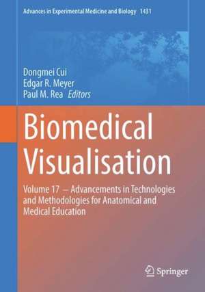
Biomedical Visualisation: Volume 17 ‒ Advancements in Technologies and Methodologies for Anatomical and Medical Education: Advances in Experimental Medicine and Biology, cartea 1431
Editat de Dongmei Cui, Edgar R. Meyer, Paul M. Reaen Limba Engleză Hardback – 31 aug 2023
This edited book explores advances in anatomical sciences education, such as teaching methods, integration of systems-based components, course design and implementation, assessments, effective learning strategies in and outside the learning environment, and novel approaches to active learning in and outside the laboratory and classroom. Many of these advances involve computer-based technologies. These technologies include virtual reality, augmented reality, mixed reality, digital dissection tables, digital anatomy apps, three-dimensional (3D) printed models, imaging and 3D reconstruction, virtual microscopy, online teaching platforms, table computers and video recording devices, software programs, and other innovations. Any of these devices and modalities can be used to develop large-class practical guides, small-group tutorials, peer teaching and assessment sessions, and various products and pathways for guided and self-directed learning.
The reader will be able to explore useful information pertaining to a variety of topics incorporating these advances in anatomical sciences education. The book will begin with the exploration of a novel approach to teaching dissection-based anatomy in the context of organ systems and functional compartments, and it will continue with topics ranging from teaching methods and instructional strategies to developing content and guides for selecting effective visualization technologies, especially in lieu of the recent and residual effects of the COVID-19 pandemic. Overall, the book covers several anatomical disciplines, including microscopic anatomy/histology, developmentalanatomy/embryology, gross anatomy, neuroanatomy, radiological imaging, and integrations of clinical correlations.
Din seria Advances in Experimental Medicine and Biology
- 9%
 Preț: 719.56 lei
 - 5%
 Preț: 717.20 lei
 - 20%
 Preț: 691.93 lei
 - 5%
 Preț: 715.71 lei
 - 5%
 Preț: 1113.83 lei
 - 5%
 Preț: 1031.00 lei
 - 15%
 Preț: 640.24 lei
 - 5%
 Preț: 717.00 lei
 - 5%
 Preț: 820.42 lei
 - 5%
 Preț: 717.00 lei
 - 5%
 Preț: 715.35 lei
 - 5%
 Preț: 716.28 lei
 - 5%
 Preț: 716.28 lei
 - 15%
 Preț: 641.38 lei
 - 20%
 Preț: 1161.71 lei
 - 5%
 Preț: 1170.51 lei
 - 18%
 Preț: 1119.87 lei
 - 5%
 Preț: 1288.48 lei
 - 5%
 Preț: 1164.67 lei
 - 5%
 Preț: 1101.73 lei
 - 18%
 Preț: 1123.67 lei
 - 5%
 Preț: 1435.64 lei
 - 20%
 Preț: 1044.10 lei
 - 18%
 Preț: 946.39 lei
 - 5%
 Preț: 292.57 lei
 - 18%
 Preț: 957.62 lei
 - 18%
 Preț: 1235.76 lei
 - 5%
 Preț: 1231.55 lei
 - 5%
 Preț: 1292.30 lei
 - 5%
 Preț: 1102.10 lei
 - 18%
 Preț: 1132.81 lei
 - 5%
 Preț: 1165.19 lei
 - 5%
 Preț: 1418.48 lei
 - 5%
 Preț: 1305.63 lei
 - 18%
 Preț: 1417.72 lei
 - 18%
 Preț: 1412.99 lei
 - 24%
 Preț: 806.15 lei
 - 18%
 Preț: 1243.29 lei
 - 5%
 Preț: 1429.44 lei
 - 5%
 Preț: 1618.70 lei
 - 5%
 Preț: 1305.12 lei
 - 18%
 Preț: 1124.92 lei
 - 5%
 Preț: 1097.54 lei
 - 15%
 Preț: 649.87 lei
 - 5%
 Preț: 1097.54 lei
 - 18%
 Preț: 945.79 lei
 - 5%
 Preț: 1123.13 lei
 - 20%
 Preț: 816.43 lei
Preț: 1291.22 lei
Preț vechi: 1359.17 lei
-5% Nou
Puncte Express: 1937
Preț estimativ în valută:
247.13€ • 257.00$ • 206.80£
247.13€ • 257.00$ • 206.80£
Carte tipărită la comandă
Livrare economică 15-29 martie
Preluare comenzi: 021 569.72.76
Specificații
ISBN-13: 9783031367267
ISBN-10: 303136726X
Pagini: 212
Ilustrații: X, 212 p. 77 illus., 74 illus. in color.
Dimensiuni: 178 x 254 mm
Greutate: 0.61 kg
Ediția:1st ed. 2023
Editura: Springer International Publishing
Colecția Springer
Seria Advances in Experimental Medicine and Biology
Locul publicării:Cham, Switzerland
ISBN-10: 303136726X
Pagini: 212
Ilustrații: X, 212 p. 77 illus., 74 illus. in color.
Dimensiuni: 178 x 254 mm
Greutate: 0.61 kg
Ediția:1st ed. 2023
Editura: Springer International Publishing
Colecția Springer
Seria Advances in Experimental Medicine and Biology
Locul publicării:Cham, Switzerland
Cuprins
Chapter 1. Past and Current Learning and Teaching Resources and Platforms.- Chapter 2. Developing a Flipped Classroom for Clinical Anatomy: Approaches to Pre-class Recordings and a Novel Approach to In-Class Active Learning.- Chapter 3. An Overview of Traditional and Advanced Visualization Techniques Applied to Anatomical Instruction Involving Cadaveric Dissection.- Chapter 4. Technology-Enhanced Preclinical Medical Education (Anatomy, Histology and Occasionally, Biochemistry): A Practical Guide.- Chapter 5. Integration of Gross Anatomy, Histology, and Pathology in a Pre-matriculation Curriculum: A Triple-Discipline Approach.- Chapter 6. Methods for Assessing Students’ Learning of Histology: A Chronology over 50 Years!.- Chapter 7. Using Stereoscopic Virtual Presentation for Clinical Anatomy Instruction and Procedural Training in Medical Education.- Chapter 8. Creating Virtual Models and 3D Movies Using DemoMaker for Anatomical Education.- Chapter 9. Teaching Cellular Architecture: TheGlobal Status of Histology Education.
Notă biografică
Dongmei Cui M.D (hon), Ph.D. is an Associate Professor at the University of Mississippi Medical Center. She led the development of 3D Virtual Anatomy Research Laboratory and mentors undergraduate students, graduate students (MS & PhD) and medical students conducting 3D educational research at the UMMC. Her educational research philosophy is to develop innovative 3D teaching tools to facilitate the acquisition of anatomical knowledge. The educational research papers from her laboratory have been published in several journals, one of them was selected as the cover page for Anatomical Science Education, and another was featured in the News Letter of the American Association of Anatomists (AAA). She was the lead author of the textbook titled "Atlas of Histology with Functional and Clinical Correlations." This textbook was ranked as one of the few leading histology textbooks by Doody’s Core Titles library referral service in 2012 and 2016. The textbook has been translated into five languages (Spanish, Italian, Indonesian, complex Chinese and Turkish editions) and is currently used for teaching histology worldwide. Her “Histology Flash Cards with Clinical Correlations” was very popular among students. She is also the lead author of the recent textbook "Histology from a Clinical Perspective". Those works were published by Wolters Kluwer/Lippincott Williams and Wilkins. She has participated in national and international meetings as a presenter and invited speaker. She also helped to organize symposium for the international meetings and was chair/co-chair and speaker at the 18th and 19th Congress of the International Federation of Associations of Anatomists (IFAA), and 25th International Symposia on Morphological Sciences (ISMS). Dr. Cui is a member of the American Association of Anatomists and served on the editorial board of Journal of Anatomical Sciences Education (2018-2022). She is a leader for International Federation of Associations ofAnatomists Task Force to develop Core Syllabus for the Histology in Medical Curriculum. She teaches Histology and Cell Biology to medical students and graduate students. She teaches Microscopic Anatomy to dental students for many years. She has developed an integrated curriculum and has served as Course Director for pre-matriculation course and currently a course director for Medical Review of Histology/with Clinical Correlation course and Course Director for Dental Histology (Human Microscopic and Development Anatomy).
Edgar R. Meyer, M.A.T., Ph.D., is an Assistant Professor with an appointment in the School of Medicine, Department of Advanced Biomedical Education at the University of Mississippi Medical Center (UMMC). He also serves as the Director of the Master of Science in Biomedical Sciences (M.S.-BMS) Program in the School of Graduate Studies in the Health Sciences at UMMC. In this role, he engages in one-on-one advising with graduate students intending to enroll in health professional programs to pursue careers in the health sciences. His research interests revolve around virtual anatomy; diversity, equity, and inclusion; and outreach endeavors that serve K-12 student and teacher populations. He has experience teaching embryology, histology, gross anatomy, and/or neuroanatomy to medical, dental, physician associate, nurse anesthesia, and graduate students.
Prof. Paul M. Rea is Professor of Digital and Anatomical Education at the University of Glasgow. He is Director of Innovation, Engagement and Enterprise within the School of Medicine, Dentistry and Nursing. He is also a Senate Assessor for Student Conduct, Council Member on Senate and coordinates the day-to-day running of the Body Donor Program and is a Licensed Teacher of Anatomy, licensed by the Scottish Parliament.
He is qualified with a medical degree (MBChB), a MSc (by research) in craniofacial anatomy/surgery, a PhD in neuroscience, the Diploma in Forensic Medical Science (DipFMS), and an MEd with Merit (Learning and Teaching in Higher Education). He is a Senior Fellow of the Higher Education Academy, Fellow of the Institute of Medical Illustrators (MIMI) and a registered medical illustrator with the Academy for Healthcare Science.
Paul has published widely and presented at many national and international meetings, including invited talks. He has been the lead Editor for Biomedical Visualiz(s)ation over 15 published volumes and is the founding editor for this book series. This has resulted in almost over 110,000 downloads across these volumes, with contributions from over 450 different authors, across approximately 100 institutions from 23 countries across the globe. It has over 500 citations from these volumes. He is Associate Editor for the European Journal of Anatomy and has reviewed for 25 different journals/publishers. He is the Public Engagement and Outreachlead for anatomy coordinating collaborative projects with the Glasgow Science Centre, NHS and Royal College of Physicians and Surgeons of Glasgow. Paul is also a STEM ambassador and has visited numerous schools to undertake outreach work. His research involves a long-standing strategic partnership with the School of Simulation and Visualisation The Glasgow School of Art. This has led to multi-million-pound investment in creating world leading 3D digital datasets to be used in undergraduate and postgraduate teaching to enhance learning and assessment. This successful collaboration resulted in the creation of the world’s first taught MSc Medical Visualisation and Human Anatomy combining anatomy and digital technologies, for which Paul was the Founding Director having managed this for 12 years. The Institute of Medical Illustrators also accredits this postgraduate degree. Paul has led college-wide, industry, multi-institutional and NHS research linked projects for students.
Textul de pe ultima copertă
Curricula in the health sciences have undergone significant change and reform in recent years. The time allocated to anatomical education in medical, osteopathic medical, and other health professional programs has largely decreased. As a result, educators are seeking effective teaching tools and useful technology in their classroom learning.
The reader will be able to explore useful information pertaining to a variety of topics incorporating these advances in anatomical sciences education. The book will begin with the exploration of a novel approach to teaching dissection-based anatomy in the context of organ systems and functional compartments, and it will continue with topics ranging from teaching methods and instructional strategies to developing content and guides for selecting effective visualization technologies, especially in lieu of the recent and residual effects of the COVID-19 pandemic. Overall, the book covers several anatomical disciplines, including microscopic anatomy/histology, developmental anatomy/embryology, gross anatomy, neuroanatomy, radiological imaging, and integrations of clinical correlations.
This edited book explores advances in anatomical sciences education, such as teaching methods, integration of systems-based components, course design and implementation, assessments, effective learning strategies in and outside the learning environment, and novel approaches to active learning in and outside the laboratory and classroom. Many of these advances involve computer-based technologies. These technologies include virtual reality, augmented reality, mixed reality, digital dissection tables, digital anatomy apps, three-dimensional (3D) printed models, imaging and 3D reconstruction, virtual microscopy, online teaching platforms, table computers and video recording devices, software programs, and other innovations. Any of these devices and modalities can be used to develop large-class practical guides, small-group tutorials, peer teaching and assessment sessions, and various products and pathways for guided and self-directed learning.
The reader will be able to explore useful information pertaining to a variety of topics incorporating these advances in anatomical sciences education. The book will begin with the exploration of a novel approach to teaching dissection-based anatomy in the context of organ systems and functional compartments, and it will continue with topics ranging from teaching methods and instructional strategies to developing content and guides for selecting effective visualization technologies, especially in lieu of the recent and residual effects of the COVID-19 pandemic. Overall, the book covers several anatomical disciplines, including microscopic anatomy/histology, developmental anatomy/embryology, gross anatomy, neuroanatomy, radiological imaging, and integrations of clinical correlations.
Caracteristici
Compares digital technologies used for virtual anatomy teaching Provides a guide for step-by-step incorporation of technology into biomedical education Describes how to use audience response technology and other forms of assessment in the learning environment
