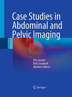
Case Studies in Abdominal and Pelvic Imaging
Autor Rita Joarder, Neil Crundwell, Matthew Gibsonen Limba Engleză Paperback – 31 aug 2014
Compiled by experts in the field, Case Studies in Abdominal and Pelvic Imaging uses the most up-to-date and high quality images, including plain films, CT scans, MRI scans and the occasional nuclear medicine image where relevant.
Each case is presented in a pedagogical style, with 1-4 images and accompanying questions, followed by answers and further relevant images. This is then augmented by an explanation of the imaging and key teaching points with references for further reading, making this book a valuable learning guide in an accessible form.
| Toate formatele și edițiile | Preț | Express |
|---|---|---|
| Paperback (1) | 879.02 lei 38-44 zile | |
| SPRINGER LONDON – 31 aug 2014 | 879.02 lei 38-44 zile | |
| Hardback (1) | 681.03 lei 38-44 zile | |
| SPRINGER LONDON – 29 mai 2011 | 681.03 lei 38-44 zile |
Preț: 879.02 lei
Preț vechi: 925.28 lei
-5% Nou
Puncte Express: 1319
Preț estimativ în valută:
168.22€ • 172.27$ • 139.93£
168.22€ • 172.27$ • 139.93£
Carte tipărită la comandă
Livrare economică 14-20 martie
Preluare comenzi: 021 569.72.76
Specificații
ISBN-13: 9781447161615
ISBN-10: 1447161610
Pagini: 316
Ilustrații: XIV, 301 p.
Dimensiuni: 193 x 260 x 17 mm
Ediția:2011
Editura: SPRINGER LONDON
Colecția Springer
Locul publicării:London, United Kingdom
ISBN-10: 1447161610
Pagini: 316
Ilustrații: XIV, 301 p.
Dimensiuni: 193 x 260 x 17 mm
Ediția:2011
Editura: SPRINGER LONDON
Colecția Springer
Locul publicării:London, United Kingdom
Public țintă
Professional/practitionerCuprins
Case 1:- Case 2:- Case 3:- Case 4:- Case 5:- Case 6:- Case 7:- Case 8:- Case 9:- Case 10:- Case 11:- Case 12 :- Case 13:- Case 14:- Case 15:- Case 16:- Case 17:- Case 18:- Case 19:- Case 20:- Case 21:- Case 22:- Case 23:- Case 24:- Case 25:- Case 26:- Case 27:- Case 28:- Case 29 :- Case 30:- Case 31:- Case 32:- Case 33:-Case 34:- Case 35:- Case 36:- Case 37:- Case 38:- Case 39:- Case 40:- Case 41:- Case 42:- Case 43:- Case 44:- Case 45:- Case 46:- Case 47:- Case 48:- Case 49:- Case 50:- Case 51:- Case 52:- Case 53:- Case 54:- Case 55:- Case 56:- Case 57:- Case 58:- Case 59:- Case 60:- Case 61:- Case 62:- Case 63:- Case 64:- Case 65:- Case 66:- Case 67:- Case 68:- Case 69:- Case 70:- Case 71:- Case 72:- Case 73:- Case 74:- Case 75:- Case 76:- Case 77:- Case 78:- Case 79:- Case 80:- Case 81:- Case 82:- Case 83:- Case 84:- Case 85:- Case 86:- Case 87:- Case 88:- Case 89:- Case 90:- Case 91:- Case 92:- Case 93:- Case 94:- Case 95:- Case 96:- Case 97:- Case 98:- Case 99:- Case 100
Recenzii
From the reviews:
“The book is primarily structured as a case-based discussion of 100 cases constituting a balanced blend of conditions encountered routinely in day-to-day practice as well as rare disease conditions. … This compilation of cases is a good teaching resource for the authors’ intended audience, particularly the radiology residents, because it exposes them to simple and complicated cases generally encountered in a busy practice. … In summary, the book accomplishes its desired objective and certainly meets the expectations of residents, fellows, and practicing radiologists … .” (Avinash Kambadakone, Radiology, Vol. 364 (2), August, 2012)
“The book is primarily structured as a case-based discussion of 100 cases constituting a balanced blend of conditions encountered routinely in day-to-day practice as well as rare disease conditions. … This compilation of cases is a good teaching resource for the authors’ intended audience, particularly the radiology residents, because it exposes them to simple and complicated cases generally encountered in a busy practice. … In summary, the book accomplishes its desired objective and certainly meets the expectations of residents, fellows, and practicing radiologists … .” (Avinash Kambadakone, Radiology, Vol. 364 (2), August, 2012)
Notă biografică
Rita Joarder, BSc.MBBS, FRCP, FRCR - Consultant Radiologist, Conquest Hospital in Hastings Neil Crundwell, MRCP FRCR - Consultant Radiologist, Conquest Hospital in Hastings Matthew Gibson, BMedSci, BMBS, MRCP, FRCR – Consultant Radiologist, Royal Berkshire Hospital, Reading
Textul de pe ultima copertă
Case Studies in Abdominal and Pelvic Imaging presents 100 case studies, covering both common every-day conditions of the abdomen and pelvis, as well as less common cases that junior doctors and radiologists in training should be aware of. Compiled by experts in the field, Case Studies in Abdominal and Pelvic Imaging uses the most up-to-date and high quality images, including plain films, CT scans, MRI scans and the occasional nuclear medicine image where relevant. Each case is presented in a pedagogical style, with 1-4 images and accompanying questions, followed by answers and further relevant images.This is then augmented by an explanation of the imaging and key teaching points with references for further reading, making this book a valuable learning guide in an accessible form.
Caracteristici
Including 2 to 4 images per case, the reader is provided with a broad coverage of the particular chest condition High quality up-to-date relevant images ensure that the case study images match the quality of those used in the hospital Key points of each case facilitates learning how to diagnose the various chest conditions A good mix of cases is presented, so that the reader enjoys a broad survey of the chest conditions they may encounter Includes supplementary material: sn.pub/extras
