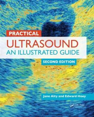
Practical Ultrasound: An Illustrated Guide, Second Edition
Autor Jane Alty, Edward Hoey, Michael Westonen Limba Engleză Paperback – 21 aug 2013
See What’s New in the Second Edition:
- New chapters on breast, musculoskeletal, and FAST (focused assessment with sonography in trauma) ultrasonography
- Revisions to original chapters incorporating up-to-date techniques and protocols
Organized according to anatomical site, the chapters include a review section on useful anatomy, scan protocol presented step by step, and a section on common pathology. Maintaining the popular format of the previous edition, each chapter contains examples of common and clinically relevant pathologies and notes on the salient features of these conditions.
The authors’ precise approach puts an immense amount of knowledge within easy reach, making it an ideal aid for learning the practicalities of ultrasound.
| Toate formatele și edițiile | Preț | Express |
|---|---|---|
| Paperback (2) | 510.59 lei 3-5 săpt. | +21.38 lei 5-11 zile |
| CRC Press – 13 noi 2024 | 510.59 lei 3-5 săpt. | +21.38 lei 5-11 zile |
| CRC Press – 21 aug 2013 | 539.84 lei 3-5 săpt. | +41.48 lei 5-11 zile |
| Hardback (1) | 1264.81 lei 6-8 săpt. | |
| CRC Press – 13 noi 2024 | 1264.81 lei 6-8 săpt. |
Preț: 539.84 lei
Preț vechi: 568.25 lei
-5% Nou
103.31€ • 107.46$ • 85.29£
Carte disponibilă
Livrare economică 24 martie-07 aprilie
Livrare express 08-14 martie pentru 51.47 lei
Specificații
ISBN-10: 1444168290
Pagini: 296
Ilustrații: 700 colour illustrations, 100 colour illustrations, 600 colour line drawings
Dimensiuni: 216 x 270 x 15 mm
Greutate: 1 kg
Ediția:Revizuită
Editura: CRC Press
Colecția CRC Press
Public țintă
Professional ReferenceCuprins
General principles of ultrasound scanning
Guide to using the ultrasound machine
Abdomen
Renal, including renal transplant
Abdominal aorta
Liver transplant
Testes
Lower limb veins
Carotid Doppler
Female pelvis
Early pregnancy
Thyroid
Focused assessment with sonography in trauma (FAST)
Breast
Muscoloskeletal
Index
Notă biografică
Jane Alty MB BChir MA MRCP, Specialist Registrar, St James's University Hospital, Leeds, UK
Edward Hoey MB BCh BAO MRCP FRCR, Specialist Registrar, St James's University Hospital, Leeds, UK
Recenzii
—Henry C Irving FRCR, Consultant Radiologist, St James’s University Hospital, Leeds, UK; Ex-Treasurer of The Royal College of Radiologists
"The authors are to be congratulated for their efforts. … Not only are you on the brink of discovering a wonderful world of non-invasive imaging, but also your pathway to achieving the competence that you desire will be made considerably easier by this book."
—Henry C. Irving FRCR, Consultant Radiologist, St James’s University Hospital, Leeds, UK; Ex-Treasurer of The Royal College of Radiologists
"The authors then walk you gently through the important steps involved in each scan, guiding the way with useful diagrams indicating probe positions and corresponding expected views of the underlying anatomy. I found these ‘templates’ for each scan incredibly useful … This book sets out to give the reader everything they need to know to become confident at ultrasound scanning."
—Jim Hare, Mersey School of Radiology, University of Liverpool, UK
Descriere
In the hands of a skilled operator, ultrasound scanning is a simple and easy procedure. However, reaching that level of proficiency can be a long and tedious process. Commended by the British Medical Association, Practical Ultrasound, Second Edition focuses on the scans regularly encountered in a busy ultrasound department and provides everything practitioners need to know to become competent and skilled in scanning.
See What’s New in the Second Edition:
- New chapters on breast, musculoskeletal, and FAST (focused assessment with sonography in trauma) ultrasonography
- Revisions to original chapters incorporating up-to-date techniques and protocols
Beginning with the general principles of ultrasound scanning and a guide to using the ultrasound machine, the book provides step-by-step instructions on how to perform scans supplemented by high-quality images and handy tips.
Organized according to anatomical site, the chapters include a review section on useful anatomy, scan protocol presented step by step, and a section on common pathology. Maintaining the popular format of the previous edition, each chapter contains examples of common and clinically relevant pathologies and notes on the salient features of these conditions.
The authors’ precise approach puts an immense amount of knowledge within easy reach, making it an ideal aid for learning the practicalities of ultrasound.
