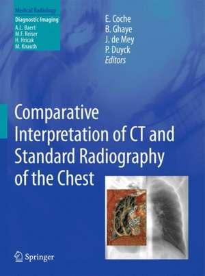
Comparative Interpretation of CT and Standard Radiography of the Chest: Medical Radiology
Editat de Emmanuel E. Coche, Benoit Ghaye, Johan de Mey, Philippe Duycken Limba Engleză Paperback – 26 mar 2014
| Toate formatele și edițiile | Preț | Express |
|---|---|---|
| Paperback (1) | 1612.60 lei 38-44 zile | |
| Springer Berlin, Heidelberg – 26 mar 2014 | 1612.60 lei 38-44 zile | |
| Hardback (1) | 1450.84 lei 3-5 săpt. | |
| Springer Berlin, Heidelberg – 8 noi 2010 | 1450.84 lei 3-5 săpt. |
Din seria Medical Radiology
- 5%
 Preț: 1108.87 lei
 - 5%
 Preț: 349.24 lei
 - 5%
 Preț: 1308.02 lei
 - 5%
 Preț: 1308.74 lei
 - 5%
 Preț: 720.68 lei
 - 5%
 Preț: 717.20 lei
 - 5%
 Preț: 1626.03 lei
 - 5%
 Preț: 1618.70 lei
 - 5%
 Preț: 802.21 lei
 - 5%
 Preț: 1130.07 lei
 - 5%
 Preț: 1116.00 lei
 - 5%
 Preț: 1953.34 lei
 - 5%
 Preț: 783.04 lei
 - 5%
 Preț: 1105.61 lei
 - 5%
 Preț: 794.00 lei
 - 5%
 Preț: 1101.21 lei
 - 5%
 Preț: 821.19 lei
 - 5%
 Preț: 1420.29 lei
 - 5%
 Preț: 743.16 lei
 - 5%
 Preț: 906.63 lei
 - 5%
 Preț: 1313.75 lei
 - 5%
 Preț: 1858.30 lei
 - 5%
 Preț: 1306.73 lei
 - 5%
 Preț: 1113.11 lei
 - 5%
 Preț: 1462.37 lei
 - 5%
 Preț: 1301.44 lei
 - 5%
 Preț: 975.17 lei
 - 5%
 Preț: 1122.58 lei
 - 5%
 Preț: 1986.27 lei
 - 5%
 Preț: 1126.82 lei
 - 5%
 Preț: 718.46 lei
 - 5%
 Preț: 1450.84 lei
 - 5%
 Preț: 1298.14 lei
 - 5%
 Preț: 1110.32 lei
 - 5%
 Preț: 1184.42 lei
 - 5%
 Preț: 1113.99 lei
 - 5%
 Preț: 1435.85 lei
 - 5%
 Preț: 663.23 lei
 - 5%
 Preț: 1605.11 lei
 - 5%
 Preț: 731.07 lei
 - 5%
 Preț: 733.09 lei
 - 5%
 Preț: 1124.07 lei
 - 5%
 Preț: 383.93 lei
 - 5%
 Preț: 1106.69 lei
 - 5%
 Preț: 982.50 lei
 - 5%
 Preț: 1317.17 lei
 - 5%
 Preț: 1437.67 lei
 - 5%
 Preț: 1307.85 lei
 - 5%
 Preț: 1950.60 lei
Preț: 1612.60 lei
Preț vechi: 1697.47 lei
-5% Nou
Puncte Express: 2419
Preț estimativ în valută:
308.61€ • 335.10$ • 259.23£
308.61€ • 335.10$ • 259.23£
Carte tipărită la comandă
Livrare economică 18-24 aprilie
Preluare comenzi: 021 569.72.76
Specificații
ISBN-13: 9783642265891
ISBN-10: 3642265898
Pagini: 492
Ilustrații: XII, 480 p.
Dimensiuni: 193 x 260 x 26 mm
Greutate: 1 kg
Ediția:2011
Editura: Springer Berlin, Heidelberg
Colecția Springer
Seriile Medical Radiology, Diagnostic Imaging
Locul publicării:Berlin, Heidelberg, Germany
ISBN-10: 3642265898
Pagini: 492
Ilustrații: XII, 480 p.
Dimensiuni: 193 x 260 x 26 mm
Greutate: 1 kg
Ediția:2011
Editura: Springer Berlin, Heidelberg
Colecția Springer
Seriile Medical Radiology, Diagnostic Imaging
Locul publicării:Berlin, Heidelberg, Germany
Public țintă
Professional/practitionerCuprins
Introduction: The Remaining Indications of Chest Radiography in Clinics.- Difficulties for Chest Radiography Interpretation.- Technical and Practical Aspects for CT Reconstruction and Image Comparison: The use of Isotropic Imaging and CT Reconstructions.- The Use of PACS, Tips and Tricks for Image Comparison.- Semeiology of Normal Variants and Diseased Chest: The Mediastinum.- The Heart.- The Hilae and Pulmonary Vessels.- The Lung Parenchyma.- The Respiratory Tract.- The Pleura.- The Diaphragm.- The Chest Wall.- Selected Diseases With Peculiar Aspect on Chest Radiography: COPD.- Missed Lung Lesions.- Lung Atelectasis.- Lung cancer.-Pulmonary Embolism and Pulmonary Hypertension.- Chest Trauma.- Subject Index.
Textul de pe ultima copertă
Standard radiography of the chest remains one of the most widely used imaging modalities but it can be difficult to interpret. The possibility of producing cross-sectional, reformatted 2D and 3D images with CT makes this technique an ideal tool for reinterpreting standard radiography of the chest. The aim of this book is to provide a comprehensive overview of chest radiography interpretation by means of a side-by-side comparison between chest radiographs and CT images. Introductory chapters address the indications for and difficulties of chest radiography as well as the technical and practical aspects of CT reconstruction and image comparison. Thereafter, the radiographic and CT presentations of both anatomical variants and the diseased chest are illustrated and discussed by renowned experts in thoracic imaging. Individual chapters are devoted to the imaging features of selected common diseases and disorders, including COPD, lung cancer, pulmonary embolism and hypertension, atelectasis and chest trauma. The book is complemented by online extra material which provides many further educational examples.
.
.
Caracteristici
Uniquely, this book provides a comprehensive overview of chest radiography interpretation by means of a side-by-side comparison between chest radiographs and CT images The radiographic and CT presentations of both anatomical variants and a wide range of diseases and disorders are illustrated and discussed by renowned experts in the field Online extra material provides many further educational examples Includes supplementary material: sn.pub/extras
