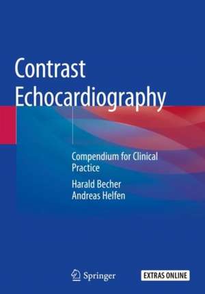
Contrast Echocardiography: Compendium for Clinical Practice
Autor Harald Becher, Andreas Helfenen Limba Engleză Paperback – 15 aug 2020
Contrast Echocardiography: Compendium for Clinical Practice comprehensively covers all aspects of the clinical use of contrast echocardiography and has been written by two cardiologists who share their experience from their high volume echo laboratories. One of the authors has been a member of both the ASE guidelines and EACVI recommendation writing groups. It is therefore, a critical text for echocardiographers and sonographers who perform echocardiography.
| Toate formatele și edițiile | Preț | Express |
|---|---|---|
| Paperback (1) | 569.75 lei 38-45 zile | |
| Springer International Publishing – 15 aug 2020 | 569.75 lei 38-45 zile | |
| Hardback (1) | 856.16 lei 3-5 săpt. | |
| Springer International Publishing – 17 iul 2019 | 856.16 lei 3-5 săpt. |
Preț: 569.75 lei
Preț vechi: 599.74 lei
-5% Nou
Puncte Express: 855
Preț estimativ în valută:
109.02€ • 113.83$ • 90.23£
109.02€ • 113.83$ • 90.23£
Carte tipărită la comandă
Livrare economică 31 martie-07 aprilie
Preluare comenzi: 021 569.72.76
Specificații
ISBN-13: 9783030159641
ISBN-10: 3030159647
Pagini: 333
Ilustrații: XXX, 333 p. 434 illus., 422 illus. in color.
Dimensiuni: 178 x 254 mm
Ediția:1st ed. 2019
Editura: Springer International Publishing
Colecția Springer
Locul publicării:Cham, Switzerland
ISBN-10: 3030159647
Pagini: 333
Ilustrații: XXX, 333 p. 434 illus., 422 illus. in color.
Dimensiuni: 178 x 254 mm
Ediția:1st ed. 2019
Editura: Springer International Publishing
Colecția Springer
Locul publicării:Cham, Switzerland
Cuprins
Introduction.- Agitated saline.- Indications.- Preparation of right heart contrast agents.- Side effects.- Gelatine-preparations.- Left heart contrast agents.- Indications.- Contraindications.- Properties of left heart contrast agents.- Side effects: diagnosis and treatment.- Metabolism of contrast agents.- Preparing the contrast agent the patient for contrast injection.- Preparing the patient.- Echocardiography scanner – setting for contrast echocardiography.- Agitated saline.- left heart contrast agents.- Infusion pump for contrast echocardiography.- Indications.- Operation of the pump.- Video-Guide.- References.- LV-Function and LV disease.- LV-Volumes und Function in 2D-und 3D-Echocardiography.- Measurement of LV-Volumes und LV-Function according current guidelines.- When is accurate measurement clinically relevant?.- Limitations of non-contrast 2D- und 3D-Echocardiography.- Advantages of Contrast Echocardiography for measurement of LV volumes.- Indications for Contrast Echocardiography.- Practice of 2D Contrast Echocardiography.- Recordings.- Criteria for adequate recordings.- Step by step analysis, how to avoid pitfalls.- Normal values.- EF-measurements in cardio-oncology.- EF measurements for ICD,CRT.- LV Volumes und EF in valvular heart disease.- EF and LV volumes in patients with heart failure.- Regional LV wall motion.- Practice of 3D Contrast Echocardiography.- Indications for using contrast agents.- 3D contrast echo recordings: how to perform/trouble shooting.- Analysis/How to avoid.- Normal values .- LV-Myocardial disease and masses.- Cardiomyopathies.- LV aneurysm.- LV masses/thrombi.- At a glance.- Video-Guide.- References.- Transesophageal Contrast Echocardiography.- Anatomy of left atrial appendage (LAA).- What is relevant to the physician?.- LAA shapes: chicken wing and non-chicken wing.- What causes LAA thrombi?.- LAA thrombi basics.- Diagnostic criteria of LAA thrombi.- Conclusion.- Echocardiography scanner settings.- General principles for adequate settings.- Analysis of contrast TEE recordings.- Conclusions.- Vivid 7 dimension settings.- Vivid E9 XD clear settings.- Philips IE 33 settings.- LAA thrombi – case studies.- Variant 1.- Variant 2.- Variant 3.- Variant 4.- Spontaneous echo contrast and thrombi.- Exclusion of thrombi using contrast echocardiography.- Pericardial effusion and contrast echocardiography.- LAA occlude.- Aortic dissection.- Myxoma.- Conclusion.- Video-Guide.- References.- Coronary sonography.- Basics of coronary sonography.- Introduction.- Physiology and pathophysiology of myocardial perfusion.- Normal flow signals of coronary arteries.- Pathological flow recordings.- Coronary flow reserve.- Principle of coronary sonography.- Practice of coronary sonography.- Left coronary artery.- Left main stem.- Circumflex artery.- Right coronary artery.- Synopsis of typical scan planes.- Bypass grafts.- Coronary anomalies.- Coronary artery stenosis.- Conclusion.- Coronary flow velocity reserve (CFR).- How to perform .- CFR und FFR.- CFR measurement in the right and circumflex arteries.- Echocardiography scanner settings.- Vivid- GE.- Philips EPIQ 7.- Siemens Acuson SC 2000.- Case study.- Conclusion.- Video-Guide.- References.- Stress echocardiography.- General principles.- Indications.- Contraindications.- Pretest probability.- Selection of the stress modality.- Echocardiographic parameters of ischemia.- Ischemic cascade.- Stress echocardiography for exclusion of significant coronary stenosis.- False positive and negative findings.- Stress echocardiography and ECG.- When to repeat stress echocardiography.- Referral to coronary angiography after stress echocardiography.- Exercise stress echocardiography in 10 steps.- Assessment of indications und contraindications.- Assessment whether physical stress is the adequate stress modality.- Patient preparation.- Required echocardiography laboratory equipment and staff.- Echocardiography scanner settings.- Preparation of the contrast agent.- Exercise protocol: mandatory recordings.- Management of the patient after finishing the test.- Assessment and reporting.- Conclusion and recommendations for patient management.- Dobutamine stress echocardiography in 10 steps.- Assessment of indications und contraindications.- Assessment whether Dobutamine stress is the adequate stress modality.- Patient preparation.- Required echocardiography laboratory equipment and staff.- Preparation of Dobutamine infusion.- Preparation of the contrast agent.- Dobutamine stress protocol: mandatory recordings.- Management of the patient after finishing the test.- Assessment and reporting.- Conclusion and recommendations for patient management.- Dobutamine stress echocardiography for assessment of myocardial viability.- At a glance.- Video guide.- References.- Multi-parametric stress echocardiography.- Rationale for multi-parametric stress echocardiography.- Basics of perfusion echocardiography.- Display of myocardial perfusion.- Flash-replenishment-method.- Triggered vs continuous recording.- Quality assessment of perfusion recordings.- Characteristics of abnormal perfusion.- Artifacts in perfusion imaging.- Echocardiography scanner settings.- Analysis of the recordings.- Adenosine.- Pharmacology.- Indications und contraindications.-Regadenoson.- Conclusion.- Practice of multi-parametric stress echocardiography.- Case studies.- Normal findings.- LAD stenosis.- Reporting.- Video-Guide.- References.- Acute coronary syndrome (ACS).- Diagnostic criteria of ACS.- Role of echocardiography.- Impact of contrast echocardiography.- Safety of contrast media in ACS.- Myocardial perfusion and risk stratification during ACS.- Case study.- Myocardial perfusion and risk stratification after ACS, stunning and no-reflow.- Case study.- Myocardial rupture, perforation of coronary arteries.- Aortic dissection/rupture.- Video-Guide.- References.- EACVI Core Syllabus.- References.
Notă biografică
Dr Andreas Helfen, MD, is a member of the “working group Cardiovascular Ultrasound” of the German Cardiac Society (GCS) and associate spokesman of the section “echocardiography” of the German Society of Ultrasound Medicine (DEGUM). He has been practising as a cardiologist since 2005 and in 2009 became the director of the echocardiography laboratory of the St.-Marien Hospital Lünen, an academic teaching hospital of the University of Münster. He performs more than 2000 echocardiographic examinations per year, including all advanced echocardiography methods. Since 2008 he organized more than 30 workshops on contrast echocardiography all over Germany and was invited as a speaker to national and international congresses. He is an approved course instructor of the German Society of Ultrasound Medicine (level III) and he has lectured in multiple stress echocardiography courses and stress refresher courses. In 2016 and in 2017 he was invited as a speaker and host to webinars of the European Association of Cardiovascular Imaging. In addition to his expertise in echocardiography Dr Helfen has gained extensive experience in interventional cardiology as well as in intensive care medicine. Dr Harald Becher, MD, PhD, former associate Professor at the University of Bonn and the chairman of the “working group Cardiovascular Ultrasound” of the German Cardiac Society (GCS) moved to Oxford, UK in 2001 and became Professor of Cardiac Ultrasound at Oxford University, and consultant cardiologist leading the echocardiography laboratory of the Oxford University Hospital. Since 2010 he is Professor of Medicine and the Heart and Stroke Foundation Chair for Cardiovascular Research at the University of Alberta, Canada. Dr Becher and his group belong to leaders in the development of contrast echocardiography and its implementation into clinical practice. Together with the physicist Peter Burns he wrote the first Handbook of Contrast Echocardiography and is one of the editors of theOxford Handbook of Echocardiography. Dr Becher is member of the writing committees of the American Society of Echocardiography and the European Association of Cardiovascular Imaging for recommendations of contrast agents in clinical practice. He is associate editor of Echocardiography and has written 10 book chapters and is the author of more than 140 scientific papers.
Textul de pe ultima copertă
This book provides a comprehensive overview of the practical aspects of contrast echocardiography. It also covers all the material in the guidelines published by the American Society of Echocardiography (ASE) in 2018 and the recommendations set out by the European Association of Cardiovascular Imaging (EACVI) in 2017. Contrast echocardiography at present is only used in 5-10% of cases, but this is expected to grow rapidly following the recommendations of the ASE and EACVI. The chapters cover the approved indications and provide practical advice on how to administer the contrast agents and how to optimize the recordings as well as how to deal with the pitfalls. The reader will find all the information on how to use contrast agents for assessment of shunts, LV volumes and function as well as myocardial diseases and masses. Detailed protocols are included for stress echocardiography and myocardial perfusion imaging. Other topics covered include the use of contrast agents for coronary sonography and transesophageal echocardiography.
Contrast Echocardiography: Compendium for Clinical Practice comprehensively covers all aspects of the clinical use of contrast echocardiography and has been written by two cardiologists who share their experience from their high volume echo laboratories. One of the authors has been a member of both the ASE guidelines and EACVI recommendation writing groups. It is therefore, a critical text for echocardiographers and sonographers who perform echocardiography.
Contrast Echocardiography: Compendium for Clinical Practice comprehensively covers all aspects of the clinical use of contrast echocardiography and has been written by two cardiologists who share their experience from their high volume echo laboratories. One of the authors has been a member of both the ASE guidelines and EACVI recommendation writing groups. It is therefore, a critical text for echocardiographers and sonographers who perform echocardiography.
Caracteristici
Includes comprehensive instruction on how to perform and interpret all clinically relevant techniques including coronary sonography and TEE with contrast agents Provides a step by step teaching guide using multiple high quality figures, movies and tables Contains checklists for the clinical practice of contrast echocardiography Features 219 videos that provide step by step technical instructions
