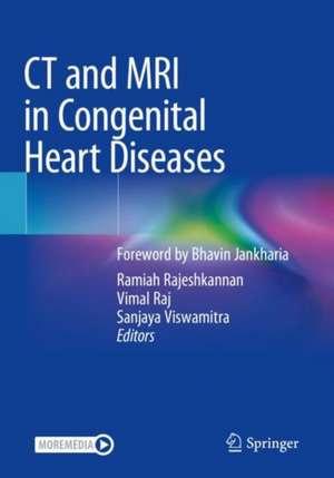
CT and MRI in Congenital Heart Diseases
Editat de Ramiah Rajeshkannan, Vimal Raj, Sanjaya Viswamitraen Limba Engleză Paperback – 19 dec 2021
Intended to provide quick and reliable access to high-quality MRI and CT images of frequently encountered congenital and structural heart abnormalities, the book offers a go-to guide for imaging physicians, helping them overcome the steep learning curve for pediatric cardiac imaging.
| Toate formatele și edițiile | Preț | Express |
|---|---|---|
| Paperback (1) | 715.93 lei 38-44 zile | |
| Springer Nature Singapore – 19 dec 2021 | 715.93 lei 38-44 zile | |
| Hardback (1) | 1138.90 lei 3-5 săpt. | |
| Springer Nature Singapore – 19 dec 2020 | 1138.90 lei 3-5 săpt. |
Preț: 715.93 lei
Preț vechi: 753.62 lei
-5% Nou
Puncte Express: 1074
Preț estimativ în valută:
136.99€ • 143.03$ • 113.38£
136.99€ • 143.03$ • 113.38£
Carte tipărită la comandă
Livrare economică 01-07 aprilie
Preluare comenzi: 021 569.72.76
Specificații
ISBN-13: 9789811567575
ISBN-10: 9811567573
Pagini: 589
Ilustrații: XIII, 589 p.
Dimensiuni: 178 x 254 mm
Greutate: 1.32 kg
Ediția:1st ed. 2021
Editura: Springer Nature Singapore
Colecția Springer
Locul publicării:Singapore, Singapore
ISBN-10: 9811567573
Pagini: 589
Ilustrații: XIII, 589 p.
Dimensiuni: 178 x 254 mm
Greutate: 1.32 kg
Ediția:1st ed. 2021
Editura: Springer Nature Singapore
Colecția Springer
Locul publicării:Singapore, Singapore
Cuprins
Part I: Basics.- CMR.- Cardiac Embryology.- Cross Sectional Imaging Atlas.- Technical Aspects of Pediatric Cardiac CT.- Scan Techniques for Pediatric Cardiac MRI.- Sequential Segmental Approach to CHD.- Part II: Imaging in Congenital Heart Disease.- Congenital Aortic Anomalies.- Vascular Rings and Slings.- Radiological Review of Coronary Artery Anomalies.- Imaging in Pulmonary Atresia with Ventricular Septal Defect.- Congenital Pulmonary Venous Anomalies.- CT and MRI of Simple Cardiovascular Shunts.- Ebstein Anomaly.- Pre and Post Operative Imaging in Tetralogy of Fallot.- Double Outlet Right Ventricle : Morphology and Function.- Pre and Post Operative Imaging in Transposition of Great Arteries.- Imaging of Single Ventricle.- CT and MR Imaging in Post Operative CHD.- Valvular Heart Disease.- Part III : Special Topics in CHD.- Techniques and Clinical Applications of Phase Contrast MRI in CHD.- Imaging of Pulmonary Hypertension in Congenital Heart Disease.- CT Versus MRI in Congenital Heart Disease.- Echocardiography for Congenital Heart Disease - Fundamental Approach.- 3D Prototyping - Technology and Applications for CHD.- Radiation issues in Pediatric Cardiac CT Imaging.
Recenzii
“This book takes you back to basic physics, anatomy and embryology and walks with you step by step to help you understand the common congenital cardiac pathologies. … The text is clear, very descriptive, easy to read and follow. Most pathologies are supported by labelled, good quality images. … I would definitely recommend reading this publication, particularly for those who have a strong interest in reporting paediatric cardiac cross-sectional imaging and building on their knowledge base in this growing area.” (Boshra Edhayr, RAD Magazine, October, 2022)
Notă biografică
Dr Ramiah Rajeshkannan (MD, DNB, PDCC) graduated from JIPMER, Puducherry University, India. He works as clinical professor at department of radiology, Amrita Institute of Medical Sciences, Kochi. He holds fellowship in cardiovascular and neuro interventional radiology from Sree Chitra Tirunal Institute of Medical Sciences, Trivandrum. He is the general secretary and founder member of Indian Association of Cardiac Imaging (IACI). The global academy of MRI has awarded him the title "ESMRMB and ISMRM certified teacher in Clinical MRI". He has many peer reviewed national and international publications.
Dr (Major). Vimal Raj (FRCR, CCT, EDM, PGDMLS) is a renowned specialist in the field of cardiothoracic imaging. He currently heads the department of radiology in Narayana Hrudayalaya, Bangalore. He has worked in three of the leading cardiothoracic centers of the world, i.e. Cambridge (Papworth Hospital), London (Royal Brompton Hospital), and Leicester (Glenfield Hospital). He has also worked with the British Army and was deployed in the war zone in Afghanistan. He has teaching interests and has published two books and multiple book chapters for radiologists and clinicians. He has published several articles in peer-reviewed journals. He also holds a patent in catheter design for performing post-mortem CT coronary angiography. He has the distinction of conducting the only Cardiac MR fellowship program in India, under the aegis of the European Society of Cardiology in India. He also holds the distinction of training more than 2000 imagers in the last three years on Cardiothoracic Imaging across the Indian subcontinent, Middle East, South Asia, Africa and Europe.
Dr. Sanjaya Viswamitra graduated from the Jawaharlal Institute of Medical Education and Research, Puducherry University, Puducherry, India in 1991. He completed a Body Imaging fellowship from Thomas Jefferson University Hospital and a Nuclear Medicine fellowship from the Hospital of the University of Pennsylvania, Philadelphia, PA. He is heading the Department of Radiology, Sri Sathya Sai Institute of Higher Medical Sciences, Bangalore. He is also a faculty at the University of Arkansas for Medical Sciences, Little Rock, AR, USA and specializes in radiology and nuclear medicine. He is the past president of the Indian Association of Cardiac Imaging and one of its founder members. He holds many Editor’s recognition awards from AJR (ARRS) and Radiology (RSNA). He has been practicing cardiac imaging since 2003 and is passionate about teaching cardiac imaging.
Textul de pe ultima copertă
This book covers the cross-sectional imaging of congenital heart diseases, and features a wealth of relevant CT and MRI images. Important details concerning anatomy, physiology, embryology and management options are discussed, and the key technical aspects of performing the imaging are explained step by step. Written by a team of respected authors, the book is richly illustrated and supplemented with access to a number of clinical videos.
Intended to provide quick and reliable access to high-quality MRI and CT images of frequently encountered congenital and structural heart abnormalities, the book offers a go-to guide for imaging physicians, helping them overcome the steep learning curve for pediatric cardiac imaging.
Caracteristici
Covers all aspects of pediatric cardiac CT and MRI Covers the latest evidence based approach to diagnosis and management of congenital heart disease Chapters are accompanied with ample images and videos to make the reading interesting Special sections on role of echo and catheter angiography and their advantages and disadvantages over cross sectional imaging is covered
