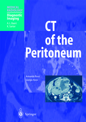
CT of the Peritoneum: Medical Radiology
Autor Armando Rossi Cuvânt înainte de A.E. Cardinale Autor Giorgio Rossi Cuvânt înainte de A.L. Baerten Limba Engleză Paperback – 6 oct 2012
| Toate formatele și edițiile | Preț | Express |
|---|---|---|
| Paperback (1) | 1329.03 lei 38-45 zile | |
| Springer Berlin, Heidelberg – 6 oct 2012 | 1329.03 lei 38-45 zile | |
| Hardback (1) | 1366.81 lei 38-45 zile | |
| Springer Berlin, Heidelberg – 20 sep 2001 | 1366.81 lei 38-45 zile |
Din seria Medical Radiology
- 5%
 Preț: 1598.56 lei
 - 5%
 Preț: 349.23 lei
 - 5%
 Preț: 1294.92 lei
 - 5%
 Preț: 1426.33 lei
 - 5%
 Preț: 708.53 lei
 - 5%
 Preț: 730.64 lei
 - 5%
 Preț: 1605.08 lei
 - 5%
 Preț: 652.06 lei
 - 5%
 Preț: 1111.02 lei
 - 5%
 Preț: 475.00 lei
 - 5%
 Preț: 1952.69 lei
 - 5%
 Preț: 1920.31 lei
 - 5%
 Preț: 1286.65 lei
 - 5%
 Preț: 1086.96 lei
 - 5%
 Preț: 706.36 lei
 - 5%
 Preț: 1411.60 lei
 - 5%
 Preț: 718.76 lei
 - 5%
 Preț: 1095.19 lei
 - 5%
 Preț: 788.71 lei
 - 5%
 Preț: 216.32 lei
 - 5%
 Preț: 1858.27 lei
 - 5%
 Preț: 377.50 lei
 - 5%
 Preț: 1094.33 lei
 - 5%
 Preț: 1437.68 lei
 - 5%
 Preț: 769.84 lei
 - 5%
 Preț: 958.70 lei
 - 5%
 Preț: 1097.17 lei
 - 5%
 Preț: 780.62 lei
 - 5%
 Preț: 1279.49 lei
 - 5%
 Preț: 1090.17 lei
 - 5%
 Preț: 705.14 lei
 - 5%
 Preț: 1276.23 lei
 - 5%
 Preț: 1103.64 lei
 - 5%
 Preț: 821.18 lei
 - 5%
 Preț: 1082.63 lei
 - 5%
 Preț: 1591.34 lei
 - 5%
 Preț: 1164.42 lei
 - 5%
 Preț: 1285.94 lei
 - 5%
 Preț: 1396.30 lei
 - 5%
 Preț: 1284.66 lei
 - 5%
 Preț: 1107.80 lei
 - 5%
 Preț: 1105.11 lei
 - 5%
 Preț: 891.35 lei
 - 5%
 Preț: 720.74 lei
 - 5%
 Preț: 1091.58 lei
 - 5%
 Preț: 1313.72 lei
 - 5%
 Preț: 1413.37 lei
 - 5%
 Preț: 1285.78 lei
 - 5%
 Preț: 1917.62 lei
Preț: 1329.03 lei
Preț vechi: 1398.98 lei
-5% Nou
Puncte Express: 1994
Preț estimativ în valută:
254.47€ • 264.98$ • 211.13£
254.47€ • 264.98$ • 211.13£
Carte tipărită la comandă
Livrare economică 10-17 februarie
Preluare comenzi: 021 569.72.76
Specificații
ISBN-13: 9783642625497
ISBN-10: 3642625495
Pagini: 432
Ilustrații: XV, 409 p. 433 illus., 6 illus. in color.
Dimensiuni: 193 x 270 x 23 mm
Ediția:Softcover reprint of the original 1st ed. 2001
Editura: Springer Berlin, Heidelberg
Colecția Springer
Seriile Medical Radiology, Diagnostic Imaging
Locul publicării:Berlin, Heidelberg, Germany
Public țintă
Professional/practitionerCuprins
Section 1: Introduction.- Normal Anatomy.- Physiology and Physiopathology of the Peritoneum.- CT Techniques.- CT Anatomy.- Section 2: Primary and Secondary Pathology of the Peritoneum.- Fluid Collections...- Acute Inflammatory Diseases.- Chronic Inflammatory Diseases.- Peritoneal and Mesenteric Trauma.- Other Nonneoplastic Pathologies.- Abdominal Herniae.- Cysts.- Primary Tumors.- Diffusion of Mahgnant Tumors of Intraperitoneal Organs to the Peritoneum, Ligaments, Mesenteries, Omentum and Lymph Nodes.
Recenzii
From the reviews:
"This book is a comprehensive, state-of-the-art review of CT in the study of anatomy and pathology of the peritoneum. … The illustrations and diagrams, as well as their reproduction, are of consistently high quality … . the book is readable and well oganised. One of the strengths of the book is the excellent chapters on anatomy. The book will be of interest for anyone interested in the diagnosis and management of mesenteric and peritoneal disease … ." (C. Valls, European Radiology, Vol. 12 (10), 2002)
"This book covers in 400 pages all thinkable aspects of the peritoneum and peritoneal cavity. … The text is exceedingly well written and the book as a whole is well organized and easy to read. There is a large number of images of excellent quality. The approach of this book is helpful to both residents and more experienced radiologists … . This book is both basic and clinical in scope and is highly recommended as an important reference in everyday radiological practice." (Hans Stridbeck, Acta Radiologica, Vol. 43 (3), 2002)
"In the foreword to this book, A. E. Cardinale describes the book as ‘splendid’. Indeed it is splendid, the illustrations and range of clinical material reflect a vast clinical experience of commonplace and rare pathology. There are beautifully produced CT sections which have been extensively researched. The text reflects the quality of the illustrations and the knowledge of the contributors. … CT departments would do themselves a significant favour in acquiring this book as a reference text." (Dr. E. M. Robertson, RAD Magazine, February, 2002)
"Though the book focuses on computed tomography (CT) of the peritoneal cavity, it also covers many aspects of abdominal imaging. … A subject index at the end of the book is extensively detailed and cross-referenced. The text … is beautifully written and carefully edited. Most of the figures are transverse CT images, but numerousCT reconstructions, standard angiograms, and diagrams are also included. All are superbly reproduced and clearly labeled. … I recommend CT of the Peritoneum to anyone interested in abdominal radiology … ." (Philip Goodman, Radiology, August, 2003)
Textul de pe ultima copertă
This superbly illustrated book, by two of the leading radiologists in Italy, is the first to be devoted entirely to computed tomography of the peritoneum. The case documentation encompasses both common and rare pathological conditions, and is the product of 20 years of painstaking research. The first part of the book is devoted to normal anatomy, physiology and physiopathology, CT techniques, and CT anatomy. The second part comprises nine chapters that systematically discuss and illustrate primary and secondary pathology of the peritoneum. The topics covered include fluid collections and effusions, acute and chronic inflammatory processes, post-traumatic lesions, hernias, cysts, primary tumors, and metastases. This book will be invaluable in improving knowledge of a topic that cannot be treated in detail in general texts on abdominal CT. Furthermore, it will be of great assistance to both radiologists and clinicians in resolving difficult and urgent diagnostic problems encountered during their everyday work.
Caracteristici
Superbly illustrated Written by two of the leading radiologists in Italy First book to be devoted entirely to computed tomography of the peritoneum The case documentation encompasses both common and rare pathological conditions, based on 20 years of research Will assist both radiologists and clinicians in resolving difficult and urgent diagnostic problems encountered during their everyday work Includes supplementary material: sn.pub/extras
