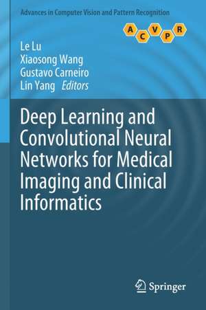
Deep Learning and Convolutional Neural Networks for Medical Imaging and Clinical Informatics: Advances in Computer Vision and Pattern Recognition
Editat de Le Lu, Xiaosong Wang, Gustavo Carneiro, Lin Yangen Limba Engleză Paperback – oct 2020
This book reviews the state of the art in deep learning approaches to high-performance robust disease detection, robust and accurate organ segmentation in medical image computing (radiological and pathological imaging modalities), and the construction and mining of large-scale radiology databases. It particularly focuses on the application of convolutional neural networks, and on recurrent neural networks like LSTM, using numerous practical examples to complement the theory.
The book’s chief features are as follows: It highlights how deep neural networks can be used to address new questions and protocols, and to tackle current challenges in medical image computing; presents a comprehensive review of the latest research and literature; and describes a range of different methods that employ deep learning for object or landmark detection tasks in 2D and 3D medical imaging. In addition, the book examines a broad selection of techniques for semantic segmentation using deep learning principles in medical imaging; introduces a novel approach to text and image deep embedding for a large-scale chest x-ray image database; and discusses how deep learning relational graphs can be used to organize a sizable collection of radiology findings from real clinical practice, allowing semantic similarity-based retrieval.
The intended reader of this edited book is a professional engineer, scientist or a graduate student who is able to comprehend general concepts of image processing, computer vision and medical image analysis. They can apply computer science and mathematical principles into problem solving practices. It may be necessary to have a certain level of familiarity with a number of more advanced subjects: image formation and enhancement, image understanding, visual recognition in medical applications, statistical learning, deep neural networks, structured prediction and image segmentation.
| Toate formatele și edițiile | Preț | Express |
|---|---|---|
| Paperback (1) | 1055.08 lei 6-8 săpt. | |
| Springer International Publishing – oct 2020 | 1055.08 lei 6-8 săpt. | |
| Hardback (1) | 1060.56 lei 6-8 săpt. | |
| Springer International Publishing – oct 2019 | 1060.56 lei 6-8 săpt. |
Din seria Advances in Computer Vision and Pattern Recognition
- 20%
 Preț: 998.36 lei
 - 20%
 Preț: 409.80 lei
 - 20%
 Preț: 654.70 lei
 - 20%
 Preț: 775.30 lei
 - 20%
 Preț: 867.15 lei
 - 20%
 Preț: 241.87 lei
 - 20%
 Preț: 659.83 lei
 - 20%
 Preț: 1084.17 lei
 - 20%
 Preț: 328.60 lei
 - 20%
 Preț: 650.08 lei
 - 20%
 Preț: 644.66 lei
 - 20%
 Preț: 652.54 lei
 - 20%
 Preț: 646.80 lei
 - 20%
 Preț: 993.60 lei
 - 20%
 Preț: 1174.92 lei
 - 20%
 Preț: 646.80 lei
 - 20%
 Preț: 672.36 lei
 - 20%
 Preț: 1166.52 lei
 - 20%
 Preț: 920.33 lei
 - 20%
 Preț: 825.78 lei
 - 20%
 Preț: 666.58 lei
 - 18%
 Preț: 953.65 lei
 - 20%
 Preț: 999.35 lei
 - 20%
 Preț: 655.02 lei
 - 20%
 Preț: 595.16 lei
 - 20%
 Preț: 647.61 lei
 - 20%
 Preț: 658.33 lei
 - 20%
 Preț: 1649.61 lei
 - 20%
 Preț: 991.46 lei
 - 20%
 Preț: 994.08 lei
 - 20%
 Preț: 1062.57 lei
 - 20%
 Preț: 985.16 lei
 - 20%
 Preț: 640.35 lei
 - 20%
 Preț: 656.03 lei
 - 18%
 Preț: 950.52 lei
 - 20%
 Preț: 652.41 lei
 - 20%
 Preț: 644.81 lei
 - 20%
 Preț: 996.40 lei
Preț: 1055.08 lei
Preț vechi: 1318.85 lei
-20% Nou
Puncte Express: 1583
Preț estimativ în valută:
201.91€ • 219.25$ • 169.61£
201.91€ • 219.25$ • 169.61£
Carte tipărită la comandă
Livrare economică 23 aprilie-07 mai
Preluare comenzi: 021 569.72.76
Specificații
ISBN-13: 9783030139711
ISBN-10: 3030139719
Pagini: 461
Ilustrații: XI, 461 p. 177 illus., 156 illus. in color.
Dimensiuni: 155 x 235 mm
Greutate: 0.72 kg
Ediția:1st ed. 2019
Editura: Springer International Publishing
Colecția Springer
Seria Advances in Computer Vision and Pattern Recognition
Locul publicării:Cham, Switzerland
Cuprins
Chapter 1. Clinical Report Guided Multi-Sieving Deep Learning for Retinal Microaneurysm Detection.- Chapter 2. Optic Disc and Cup Segmentation Based on Multi-label Deep Network for Fundus Glaucoma Screening.- Chapter 3. Thoracic Disease Identification and Localization with Limited Supervision.- Chapter 4. ChestX-ray8: Hospital-scale Chest X-ray Database and Benchmarks on Weakly-Supervised Classification and Localization of Common Thorax Diseases.- Chapter 5. TieNet: Text-Image Embedding Network for Common Thorax Disease Classification and Reporting in Chest X-rays.- Chapter 6. Deep Lesion Graph in the Wild: Relationship Learning and Organization of Significant Radiology Image Findings in a Diverse Large-scale Lesion Database.- Chapter 7. Deep Reinforcement Learning based Attention to Detect Breast Lesions from DCE-MRI.- Chapter 8. Deep Convolutional Hashing for Low Dimensional Binary Embedding of Histopathological Images.- Chapter 9. Pancreas Segmentation in CT and MRI Images via Domain Specific Network Designing and Recurrent Neural Contextual Learning.- Chapter 10. Spatial Clockwork Recurrent Neural Network for Muscle Perimysium Segmentation.- Chapter 11. Pancreas.- Chapter 12. Multi-Organ.- Chapter 13. Convolutional Invasion and Expansion Networks for Tumor Growth Prediction.- Chapter 14. Cross-Modality Synthesis in Magnetic Resonance Imaging.- Chapter 15. Image Quality Assessment for Population Cardiac MRI.- Chapter 16. Low Dose CT Image Denoising Using a Generative Adversarial Network with Wasserstein Distance and Perceptual Loss.- Chapter 17. Unsupervised Cross-Modality Domain Adaptation of ConvNets for Biomedical Image Segmentations with Adversarial Loss.- Chapter 18. Automatic Vertebra Labeling in Large-Scale Medical Images using Deep Image-to-Image Network with Message Passing and Sparsity Regularization.- Chapter 19. 3D Anisotropic Hybrid Network: Transferring Convolutional Features from 2D Images to 3D Anisotropic Volumes.- Chapter 20. Multi-Agent Learning for Robust Image Registration.- Chapter 21. Deep Learning in Magnetic Resonance Imaging of Cardiac Function.- Chapter 22. Automatic Vertebra Labeling in Large-Scale Medical Images using Deep Image-to-Image Network with Message Passing and Sparsity Regularization.- Chapter 23. Deep Learning on Functional Connectivity of Brain: Are We There Yet?.
Notă biografică
Dr. Le Lu is the Director of Ping An Technology US Research Labs, and an adjunct faculty member at Johns Hopkins University, USA.
Dr. Xiaosong Wang is a Senior Applied Research Scientist at Nvidia Corp., USA.
Dr. Gustavo Carneiro is an Associate Professor at the University of Adelaide, Australia.
Dr. Lin Yang is an Associate Professor at the University of Florida, USA.
Dr. Xiaosong Wang is a Senior Applied Research Scientist at Nvidia Corp., USA.
Dr. Gustavo Carneiro is an Associate Professor at the University of Adelaide, Australia.
Dr. Lin Yang is an Associate Professor at the University of Florida, USA.
Textul de pe ultima copertă
This book reviews the state of the art in deep learning approaches to high-performance robust disease detection, robust and accurate organ segmentation in medical image computing (radiological and pathological imaging modalities), and the construction and mining of large-scale radiology databases. It particularly focuses on the application of convolutional neural networks, and on recurrent neural networks like LSTM, using numerous practical examples to complement the theory.
The book’s chief features are as follows: It highlights how deep neural networks can be used to address new questions and protocols, and to tackle current challenges in medical image computing; presents a comprehensive review of the latest research and literature; and describes a range of different methods that employ deep learning for object or landmark detection tasks in 2D and 3D medical imaging. In addition, the book examines a broad selection of techniques for semantic segmentation using deep learning principles in medical imaging; introduces a novel approach to text and image deep embedding for a large-scale chest x-ray image database; and discusses how deep learning relational graphs can be used to organize a sizable collection of radiology findings from real clinical practice, allowing semantic similarity-based retrieval.
The intended reader of this edited book is a professional engineer, scientist or a graduate student who is able to comprehend general concepts of image processing, computer vision and medical image analysis. They can apply computer science and mathematical principles into problem solving practices. It may be necessary to have a certain level of familiarity with a number of more advanced subjects: image formation and enhancement, image understanding, visual recognition in medical applications, statistical learning, deep neural networks, structured prediction and image segmentation.
Caracteristici
Reviews the state of the art in deep learning approaches to robust disease detection, organ segmentation in medical image computing, and the construction and mining of large-scale radiology databases Particularly focuses on the application of convolutional neural networks, supporting the theory with numerous practical examples Highlights how deep neural networks can be used to address new questions and protocols, and provide novel solutions
