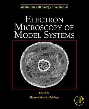
Electron Microscopy of Model Systems: Methods in Cell Biology, cartea 96
Thomas Muller-Reicherten Limba Engleză Hardback – 23 sep 2010
- Covers the preparation and analysis of model systems for biological electron microscopy
- Includes the most popular systems but also organisms that are less frequently used in cell biology
- Presents the currently most important methods for the preparation of biological specimens
- First compendium covering the various aspects of sample preparation of very diverse biological systems
Din seria Methods in Cell Biology
- 31%
 Preț: 702.75 lei
 - 31%
 Preț: 737.25 lei
 - 9%
 Preț: 734.41 lei
 - 31%
 Preț: 696.19 lei
 - 31%
 Preț: 695.58 lei
 - 32%
 Preț: 694.90 lei
 - 27%
 Preț: 702.22 lei
 - 24%
 Preț: 685.52 lei
 - 9%
 Preț: 621.87 lei
 - 31%
 Preț: 704.39 lei
 - 27%
 Preț: 745.66 lei
 - 31%
 Preț: 699.50 lei
 - 31%
 Preț: 703.51 lei
 - 27%
 Preț: 700.08 lei
 - 31%
 Preț: 706.55 lei
 - 23%
 Preț: 698.33 lei
 - 31%
 Preț: 700.08 lei
 - 31%
 Preț: 699.23 lei
 - 31%
 Preț: 749.64 lei
 - 31%
 Preț: 747.89 lei
 - 32%
 Preț: 741.01 lei
 - 9%
 Preț: 746.61 lei
 - 28%
 Preț: 697.85 lei
 - 31%
 Preț: 742.72 lei
 - 28%
 Preț: 700.51 lei
 - 32%
 Preț: 812.63 lei
 - 31%
 Preț: 818.59 lei
 - 32%
 Preț: 815.13 lei
 - 32%
 Preț: 811.43 lei
 - 32%
 Preț: 811.16 lei
 - 32%
 Preț: 790.48 lei
 - 9%
 Preț: 818.84 lei
 - 31%
 Preț: 820.31 lei
 - 33%
 Preț: 800.96 lei
 - 33%
 Preț: 800.70 lei
 - 32%
 Preț: 810.82 lei
 - 39%
 Preț: 810.62 lei
 - 29%
 Preț: 802.50 lei
 - 33%
 Preț: 799.66 lei
 - 33%
 Preț: 797.41 lei
 - 39%
 Preț: 801.98 lei
 - 39%
 Preț: 805.96 lei
 - 28%
 Preț: 814.11 lei
 - 28%
 Preț: 817.11 lei
Preț: 745.04 lei
Preț vechi: 1079.78 lei
-31% Nou
Puncte Express: 1118
Preț estimativ în valută:
142.58€ • 154.82$ • 119.77£
142.58€ • 154.82$ • 119.77£
Carte tipărită la comandă
Livrare economică 15-29 aprilie
Preluare comenzi: 021 569.72.76
Specificații
ISBN-13: 9780123810076
ISBN-10: 0123810078
Pagini: 744
Dimensiuni: 191 x 235 x 30 mm
Greutate: 1.4 kg
Editura: ELSEVIER SCIENCE
Seria Methods in Cell Biology
ISBN-10: 0123810078
Pagini: 744
Dimensiuni: 191 x 235 x 30 mm
Greutate: 1.4 kg
Editura: ELSEVIER SCIENCE
Seria Methods in Cell Biology
Public țintă
Researches, Scientist and students in Cell BiologyCuprins
Preface/Acknowledgements
Thomas Müller-Reichert
1. Electron microscopy of viruses
Michael Laue
2. Bacterial TEM: New insights from cryo-microscopy
Martin Pilhofer, Mark S. Ladinsky, Alasdair W. McDowall, and Grant Jensen*
3. Analysis of the ultrastructure of archaea by electron microscopy
Reinhard Rachel* , Carolin Meyer, Andreas Klingl, Sonja Gürster, Nadine Wasserburger, Ulf Küper, Annett Bellack, Simone Schopf, Reinhard Wirth, Harald Huber, and Gerhard Wanner
4. Chlamydomonas: Cryopreparation methods for the 3-D analysis of cellular organelles
Eileen T. O’Toole
5. Ultrastructure of the asexual blood stages of Plasmodium falciparum
Eric Hanssen* , Kenneth N Goldie, and Leann Tilley
6. Electron tomography and immunolabeling of Tetrahymena thermophila basal bodies
Thomas H. Giddings* Jr., Janet B. Meehl, Chad G. Pearson, and Mark Winey
7. Electron microscopy of Paramecium
Klaus Hausmann* and Richard D. Allen
8. Ultrastructural investigation methods for Trypanosoma brucei
Johanna L. Höög* , Eva Gluenz, Sue Vaughan, and Keith Gull
9. Dictyostelium discoideum: A model system for ultrastructural analyses of cell motility and development
Michael P. Koonce* and Ralpf Gräf
10. Towards sub-second correlative live-cell light and electron microscopy of Saccharomyces cerevisiae
Christopher Buser
11. Fission yeast: A cellular model well suited for electron microscopic investigations
Hélio D. Roque and Claude Antony*
12. High-pressure freezing and electron microscopy of Arabidopsis
Byung-Ho Kang
13. Standard and cryo-preparation of Hydra for transmission electron microscopy
Thomas W. Holstein* , Michael W. Hess, and Willi Salvenmoser
14. Electron microscopy of flatworms: Standard and cryopreparation methods
Willi Salvenmoser* , Bernhard Egger, Johannes G. Achatz, Peter Ladurner, and Michael W. Hess*
15. Three-dimensional reconstruction methods for C. elegans ultrastructure
Thomas Müller-Reichert* , Joel Mancuso, Ben Lich, and Kent McDonald*
16. Insects as model systems in cell biology
Thomas A. Keil* and R. Alexander Steinbrecht
17. Electron microscopy of amphibian model systems (Xenopus laevis and Ambystoma mexicanum)
Thomas Kurth* , Jürgen Berger, Michaela Wilsch-Bräuninger, Robert Cerny, Heinz Schwarz, Jan Löfberg, Thomas Piendl, and Hans Epperlein
18. Modern approaches for ultrastructural analysis of the zebrafish embryo
Nicole L. Schieber, Susan J. Nixon, Richard I. Webb* , and Robert G. Parton*
19. Transmission electron microscopy of cartilage and bone
Douglas R. Keene* , and Sara F. Tufa
20. Electron microscopy of the mouse central nervous system
Wiebke Möbius* , Frédérique Varoqueaux, Benjamin H. Cooper, Cordelia Imig, Torben Ruhwedel, Aiman S. Saab, and Walter Kaufmann
21. Rapidly excised and cryofixed rat tissue
Dimitri Vanhecke, Werner Graber, and Daniel Studer*
22. The actin cytoskeleton in whole mounts and sections
Guenter P. Resch* , Edit Urban, and Sonja Jacob
23. Three-dimensional cryo-electron microscopy on intermediate filaments: A critical comparison to other structural methods
Robert Kirmse, Cédric Bouchet-Marquis, Cynthia Page, and Andreas Hoenger*
24. Correlative time-lapse imaging and electron microscopy to study abscission in HeLa cells
Julien Guizetti, Jana Mäntler, Thomas Müller-Reichert* , and Daniel W. Gerlich*
25. Viral infection of cells in culture: Approaches for electron microscopy
Paul Walther* , Li Wang, Sandra Ließem, and Giada Frascaroli
26. Intracellular membrane traffic at high resolution
Jan R. T. van Weering, Edward Brown, Thomas H. Sharp, Judith Mantell, Peter J. Cullen, and Paul Verkade*
27. 3D versus 2D cell culture: Implications for electron microscopy
Michael W. Hess* , Kristian Pfaller, Hannes L. Ebner, Beate Beer, Daniel Hekl, and Thomas Seppi
28. ‘Tips and tricks’ for high-pressure freezing of model systems
Kent McDonald* , Heinz Schwarz, Thomas Müller-Reichert, Rick Webb, Christopher Buser, and Mary Morphew
