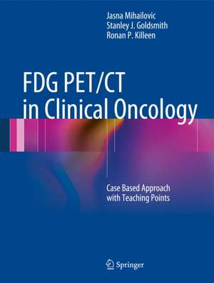
FDG PET/CT in Clinical Oncology: Case Based Approach with Teaching Points
Autor Jasna Mihailovic, Stanley J. Goldsmith, Ronan P. Killeenen Limba Engleză Hardback – 29 oct 2012
| Toate formatele și edițiile | Preț | Express |
|---|---|---|
| Paperback (1) | 694.07 lei 38-44 zile | |
| Springer Berlin, Heidelberg – 23 aug 2016 | 694.07 lei 38-44 zile | |
| Hardback (1) | 760.18 lei 22-36 zile | |
| Springer Berlin, Heidelberg – 29 oct 2012 | 760.18 lei 22-36 zile |
Preț: 760.18 lei
Preț vechi: 800.18 lei
-5% Nou
Puncte Express: 1140
Preț estimativ în valută:
145.51€ • 158.11$ • 122.30£
145.51€ • 158.11$ • 122.30£
Carte disponibilă
Livrare economică 31 martie-14 aprilie
Preluare comenzi: 021 569.72.76
Specificații
ISBN-13: 9783642298653
ISBN-10: 3642298656
Pagini: 453
Ilustrații: XIV, 453 p.
Dimensiuni: 210 x 279 x 27 mm
Greutate: 1.52 kg
Ediția:2012
Editura: Springer Berlin, Heidelberg
Colecția Springer
Locul publicării:Berlin, Heidelberg, Germany
ISBN-10: 3642298656
Pagini: 453
Ilustrații: XIV, 453 p.
Dimensiuni: 210 x 279 x 27 mm
Greutate: 1.52 kg
Ediția:2012
Editura: Springer Berlin, Heidelberg
Colecția Springer
Locul publicării:Berlin, Heidelberg, Germany
Public țintă
Professional/practitionerCuprins
PART ONE – DIAGNOSIS: Solitary pulmonary nodule/chest mass.- Unknown primary.- Unsuspected malignancy/second primary.- PART TWO – EXTENT OF DISEASE: Head & Neck.- Lung.- Breast.- Esophageal.- Colorectal.- Lymphoma.- Multiple Myeloma.- Gynecology.- Miscellaneous.- PART THREE – FOLLOW UP: Lung Ca.- Breast Ca.- Pancreas Ca.- Colorectal Ca.- Lymphoma.- Multiple Myeloma.- Thyroid Gland.- Urologic.- Miscellaneous.- PART FOUR - EVALUATION OF RESPONSE: Lymphoma.- Multiple Mueloma.- Lung.- Gastrointestinal Tumors.- Breast.- Neuroendocrine Tumor.
Recenzii
From the reviews:
“This book, intended for those seeking additional perspectives on the growing field of 18F-FDG PET/CT, uses a case-based design with teaching points. … this book will be a good guide for the nuclear medicine physician, and the authors are to be thanked for producing it.” (Hyo Jung Seo, The Journal of Nuclear Medicine, Vol. 54 (10), October, 2013)
“The authors have done a creditable job putting together 100 cases that illustrate the breadth … of current PETCT use in oncology. Individuals already experienced in PETCT reporting will find several cases of interest and educational but this book is firmly aimed at newcomers, be they nuclear medicine physicians, radiologists or clinical oncologists, and succeeds in providing an illustrated overview of FDG PETCT in oncology.” (Chirag Patel, RAD Magazine, May, 2013)
“This text, organized to reflect problems that physicians face in their daily practice, clearly demonstrates the pivotal role of FDG PET in diagnosis, staging and follow-up of patients with cancer. FDG PET/CT in Clinical Oncology is of interest mainly to residents in nuclear medicine and radiology, but it may also be of interest to practitioners working in diagnostic imaging and all clinicians who are interested in better understanding the questions that can be answered by FDG PET in daily clinical practice.” (Antonio Manna and Luigi Mansi, European Journal of Nuclear Medicine and Molecular Imaging, Vol. 40, 2013)
Textul de pe ultima copertă
FDG PET/CT has rapidly emerged as an invaluable combined imaging modality that can identify tumors on the basis of not only anatomical alterations but also metabolic activity, thus allowing the detection of lesions that would otherwise be too small to distinguish.
This book, comprising a collection of images from oncology cases, is organized according to the role of FDG PET/CT in the evaluation and management of oncology patients, and only secondarily by organ or tumor entity. In this way, it reflects the issues that clinicians actually address in their daily practice, namely: identification of an unknown or unsuspected primary; determination of the extent of disease; evaluation of response to therapy; and surveillance after response, i.e., detection of recurrent disease. In total, 100 cases involving different primary tumors are presented to illustrate findings in these different circumstances. FDG PET/CT in Clinical Oncology will be of great value to all newcomers to this field, whether medical students, radiology, nuclear medicine, or oncology fellows, or practicing physicians.
This book, comprising a collection of images from oncology cases, is organized according to the role of FDG PET/CT in the evaluation and management of oncology patients, and only secondarily by organ or tumor entity. In this way, it reflects the issues that clinicians actually address in their daily practice, namely: identification of an unknown or unsuspected primary; determination of the extent of disease; evaluation of response to therapy; and surveillance after response, i.e., detection of recurrent disease. In total, 100 cases involving different primary tumors are presented to illustrate findings in these different circumstances. FDG PET/CT in Clinical Oncology will be of great value to all newcomers to this field, whether medical students, radiology, nuclear medicine, or oncology fellows, or practicing physicians.
Caracteristici
Organized according to the role of FDG PET/CT in the evaluation and management of oncology patients 100 informative cases reflecting the issues that clinicians address in their daily practice Ideal for all newcomers to the field, whether medical students, radiology, nuclear medicine, or oncology fellows, or practicing physicians Includes supplementary material: sn.pub/extras
