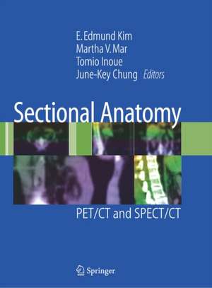
Sectional Anatomy: PET/CT and SPECT/CT
Editat de E. Edmund Kim, Martha V. Mar, Tomio Inoue, June-Key Chungen Limba Engleză Hardback – 26 noi 2007
| Toate formatele și edițiile | Preț | Express |
|---|---|---|
| Paperback (1) | 437.40 lei 43-57 zile | |
| Springer – 9 dec 2009 | 437.40 lei 43-57 zile | |
| Hardback (1) | 437.38 lei 38-44 zile | |
| Springer – 26 noi 2007 | 437.38 lei 38-44 zile |
Preț: 437.38 lei
Preț vechi: 460.39 lei
-5% Nou
Puncte Express: 656
Preț estimativ în valută:
83.69€ • 87.60$ • 69.66£
83.69€ • 87.60$ • 69.66£
Carte tipărită la comandă
Livrare economică 26 martie-01 aprilie
Preluare comenzi: 021 569.72.76
Specificații
ISBN-13: 9780387382968
ISBN-10: 0387382968
Pagini: 468
Ilustrații: XI, 468 p.
Dimensiuni: 210 x 279 x 27 mm
Greutate: 1.71 kg
Ediția:2007
Editura: Springer
Colecția Springer
Locul publicării:New York, NY, United States
ISBN-10: 0387382968
Pagini: 468
Ilustrații: XI, 468 p.
Dimensiuni: 210 x 279 x 27 mm
Greutate: 1.71 kg
Ediția:2007
Editura: Springer
Colecția Springer
Locul publicării:New York, NY, United States
Public țintă
Professional/practitionerCuprins
Normal Anatomy of PET/CT and SPECT/CT.- FDG PET/CT.- Non-FDG PET/CT.- Lymphoscintigraphy SPECT/CT.- Lung SPECT/CT.- Parathyroid SPECT/CT.- Bone SPECT/CT.- 131-I SPECT/CT.- MIBG SPECT/CT.- Gallium SPECT/CT.- Octreotide SPECT/CT.- Anatomic Variations and Artifacts of PET/CT and SPECT/CT.- PET/CT Anatomy: Variations and Artifacts.- SPECT/CT Anatomy: Variations and Artifacts.
Recenzii
From the reviews:
"This book is an image-based guide to the sectional anatomy of fusion images obtained with PET/CT and SPECT/CT scanners. ... The book is aimed primarily at clinicians who routinely interpret images and is a useful reference for most molecular imaging probes currently used with PET/CT and SPECT/CT. ... this book would be a valuable resource for anyone reading PET/CT and SPECT/CT images, providing nuclear medicine and radiology physicians, especially with a practical reference for image interpretation." (Martin Allen-Auerbach, The Journal of Nuclear Medicine, Vol. 49, 2008)
"This book is an image-based guide to the sectional anatomy of fusion images obtained with PET/CT and SPECT/CT scanners. ... The book is aimed primarily at clinicians who routinely interpret images and is a useful reference for most molecular imaging probes currently used with PET/CT and SPECT/CT. ... this book would be a valuable resource for anyone reading PET/CT and SPECT/CT images, providing nuclear medicine and radiology physicians, especially with a practical reference for image interpretation." (Martin Allen-Auerbach, The Journal of Nuclear Medicine, Vol. 49, 2008)
Caracteristici
Cutting-edge presentation of advancements in PET/CT and SPECT/CT imaging Offers guidance on the proper interpretation of PET/CT and SPECT/CT by emphasizing anatomical names and variations and artifacts Well-regarded contributors are from several top international imaging centers Beautifully illustrated, many images in color
