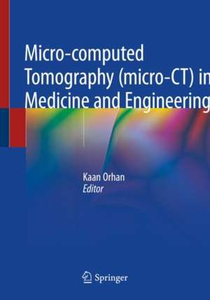
Micro-computed Tomography (micro-CT) in Medicine and Engineering
Editat de Kaan Orhanen Limba Engleză Paperback – 13 aug 2020
It particularly highlights the scanning procedure, which represents the most crucial step in micro CT, and discusses in detail the reconstruction process and the artifacts related to the scanning processes, as well as the imaging software used in analysis.
Written by international experts, the book illustrates the application of micro CT in different areas, such as dentistry, medicine, tissue engineering, aerospace engineering, geology, material engineering, civil engineering and additive manufacturing.
Covering different areas of application, the book is of interest not only to specialists in the respective fields, but also to broader audience of professionals working in the fields of imaging and analysis, as well as to students of the different disciplines.
| Toate formatele și edițiile | Preț | Express |
|---|---|---|
| Paperback (1) | 567.20 lei 38-44 zile | |
| Springer International Publishing – 13 aug 2020 | 567.20 lei 38-44 zile | |
| Hardback (1) | 854.87 lei 3-5 săpt. | |
| Springer International Publishing – 12 aug 2019 | 854.87 lei 3-5 săpt. |
Preț: 567.20 lei
Preț vechi: 597.05 lei
-5% Nou
Puncte Express: 851
Preț estimativ în valută:
108.55€ • 112.91$ • 89.61£
108.55€ • 112.91$ • 89.61£
Carte tipărită la comandă
Livrare economică 10-16 aprilie
Preluare comenzi: 021 569.72.76
Specificații
ISBN-13: 9783030166434
ISBN-10: 3030166430
Pagini: 312
Ilustrații: XII, 312 p. 212 illus., 172 illus. in color.
Dimensiuni: 178 x 254 mm
Ediția:1st ed. 2020
Editura: Springer International Publishing
Colecția Springer
Locul publicării:Cham, Switzerland
ISBN-10: 3030166430
Pagini: 312
Ilustrații: XII, 312 p. 212 illus., 172 illus. in color.
Dimensiuni: 178 x 254 mm
Ediția:1st ed. 2020
Editura: Springer International Publishing
Colecția Springer
Locul publicării:Cham, Switzerland
Cuprins
Chapter 1: Introduction to Micro-CT Imaging.- Chapter 2: X-ray Imaging: Fundamentals of X-ray.- Chapter 3: Fundamentals of Micro-CT Imaging.- Chapter 4: Artifacts in Micro-CT.- Chapter 5: Application of Bone Morphometry and Densitometry by X-ray Micro-CT to Bone Disease Models and Phenotypes.- Chapter 6: Analysis of Fracture Callus Mechanical Properties Using Micro-CT.- Chapter 7: Micro-CT in Osteoporosis Research.- Chapter 8: Micro-CT in Comparison with Histology in the Qualitative Assessment of Bone and Pathologies.- Chapter 9: Micro-CT in Artificial Tissues.- Chapter 10: Applications of Micro-CT in Soft Tissue Specimen Imaging.- Chapter 11: Applications of Micro-CT in Cardiovascular Engineering and Bio-inspired Design.- Chapter 12: Applications of Micro-CT Technology in Endodontics.- Chapter 13: Micro computed Tomography (Micro-CT) Analysis as a New Approach for Characterization of Drug Delivery Systems.- Chapter 14: Challenges in Micro-CT Characterization of Composites.- Chapter 15: Modeling and Mechanical Analysis Considerations of Structures Based on Micro-CT Data.- Chapter 16: Micro-CT Usage in Materials Science and Aerospace Engineering.- Chapter 17: X-Ray Computed Tomography Technique in Civil Engineering.- Chapter 18: Application of X-Ray Microtomography in Pyroclastic Rocks.- Chapter 19: Detection of Dispersion and Venting Quality in Plastic Composite Granules Using Micro-CT.
Notă biografică
Kaan Orhan, DDS MSc MHM PhD, BBAc is a Professor of Dentomaxillofacial Radiology at the Faculty of Dentistry, Ankara University, Turkey and a Visiting Professor at the OMFS-IMPATH Research Group, Department of Imaging and Pathology, University of Leuven, Belgium.
Dr Orhan received his degree in 1998 and completed his PhD and maxillofacial radiology residency studies in 2002 at the Osaka University Faculty of Dentistry in Osaka, Japan and Ankara University, Faculty of Dentistry. In 2004, he began his academic career at Ankara University as a consultant at the Faculty of Dentistry. He became an associate professor in 2006 and a full-time professor in 2012. From 2007 to 2010, he was the founder and chairman of the Dentomaxillofacial Radiology Department, Near East University, Cyprus.
Dr Orhan received his degree in 1998 and completed his PhD and maxillofacial radiology residency studies in 2002 at the Osaka University Faculty of Dentistry in Osaka, Japan and Ankara University, Faculty of Dentistry. In 2004, he began his academic career at Ankara University as a consultant at the Faculty of Dentistry. He became an associate professor in 2006 and a full-time professor in 2012. From 2007 to 2010, he was the founder and chairman of the Dentomaxillofacial Radiology Department, Near East University, Cyprus.
He served as chairman of the Research and Scientific Committee, European Academy of DentoMaxillofacial Radiology from 2008 to 2012, and later as Vice-President (2012-2014) and President. He is a fellow of the Japanese Board of DentoMaxillofacial Radiology, European Head and Neck Radiology Society (ESHNR), European Society of Magnetic Resonance in Medicine and Biology (ESRMB), and Turkish Magnetic Resonance Society.
Dr Orhan is an editor for several journals, including “Radiology: Open Access” and “Oral Radiology”, and a reviewer for more than 50 different journals in his field. He has authored / coauthored eight books in English and Turkish, as well as over 200 SCI international publications in peer-reviewed journals. His research interests include CT, CBCT, MRI, ultrasonography (USG), head & neck radiology, and particularly Micro-CT.
Textul de pe ultima copertă
This book focuses on applications of micro CT, CBCT and CT in medicine and engineering, comprehensively explaining the basic principles of these techniques in detail, and describing their increasing use in the imaging field.
It particularly highlights the scanning procedure, which represents the most crucial step in micro CT, and discusses in detail the reconstruction process and the artifacts related to the scanning processes, as well as the imaging software used in analysis,.
Written by international experts, the book illustrates the application of micro CT in different areas, such as dentistry, medicine, tissue engineering, aerospace engineering, geology, material engineering, civil engineering and additive manufacturing.
Covering different areas of application, the book is of interest not only to specialists in the respective fields, but also to broader audience of professionals working in the fields of imaging and analysis, as well as to students of the different disciplines.
Caracteristici
Includes numerous illustrations and sample images obtained from micro CT, CBCT and CT Presents protocols used for scanning, reconstruction and analysis Represents an invaluable tool for professionals working in the fields of application of these imaging techniques
