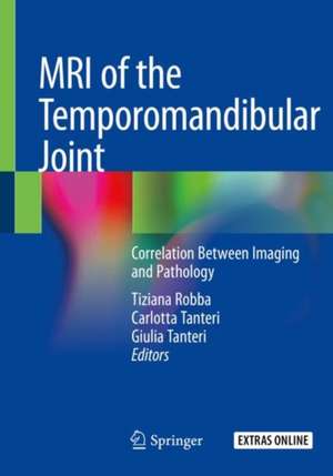
MRI of the Temporomandibular Joint: Correlation Between Imaging and Pathology
Editat de Tiziana Robba, Carlotta Tanteri, Giulia Tanterien Limba Engleză Paperback – 15 noi 2020
The authors provide essential information on TMJ anatomy, dynamics, function and dysfunction. Correlations between clinical aspects and MRI findings are discussed and guidance for the correct interpretation of results is offered. Special findings that are helpful for differential diagnosis (arthritis, osteochondroma, synovial chondromatosis) are also examined. Given its extensive and varied coverage, the book offers a valuable asset for radiologists, dentists, gnathologists, maxillofacial surgeons, orthodontists and other professionals seeking a thorough overview of the subject
| Toate formatele și edițiile | Preț | Express |
|---|---|---|
| Paperback (1) | 588.59 lei 38-44 zile | |
| Springer International Publishing – 15 noi 2020 | 588.59 lei 38-44 zile | |
| Hardback (1) | 823.79 lei 3-5 săpt. | +37.16 lei 4-10 zile |
| Springer International Publishing – 15 noi 2019 | 823.79 lei 3-5 săpt. | +37.16 lei 4-10 zile |
Preț: 588.59 lei
Preț vechi: 619.56 lei
-5% Nou
Puncte Express: 883
Preț estimativ în valută:
112.63€ • 117.89$ • 93.74£
112.63€ • 117.89$ • 93.74£
Carte tipărită la comandă
Livrare economică 27 martie-02 aprilie
Preluare comenzi: 021 569.72.76
Specificații
ISBN-13: 9783030254230
ISBN-10: 3030254232
Pagini: 238
Ilustrații: XVII, 238 p. 194 illus., 84 illus. in color.
Dimensiuni: 178 x 254 mm
Ediția:1st ed. 2020
Editura: Springer International Publishing
Colecția Springer
Locul publicării:Cham, Switzerland
ISBN-10: 3030254232
Pagini: 238
Ilustrații: XVII, 238 p. 194 illus., 84 illus. in color.
Dimensiuni: 178 x 254 mm
Ediția:1st ed. 2020
Editura: Springer International Publishing
Colecția Springer
Locul publicării:Cham, Switzerland
Cuprins
TMJ Magnetic Resonance: Technical Considerations:Principle of Physics of Magnetic Resonance Imaging TMJ.- Coils and sequences of TMJ MRI TMJ anatomy.- Normal MRI anatomy of TMJ.- TMJ dynamics.- Most common temporo-mandibular disorders:disc displacement with and without reduction hypermobility/hypomobility.- Osteoarthritis (deviations in form).- Arthritis (psoriasic,etc).- Idiopathic condylar reabsorption.- Special findings.
Notă biografică
Dr. Tiziana Robba, MD, Specialist in Diagnostic Imaging and Radiology, has been serving as a musculoskeletal radiologist at the Orthopedic Hospital and Trauma Centre of Turin, Italy (CTO – Città della Salute e della Scienza di Torino) since 2001. Her experience mainly includes traumatology, musculoskeletal and temporomandibular joint MRI, as well as musculoskeletal oncology imaging.
Dr. Carlotta Tanteri, D.D.S. M.Sc. studied Dentistry at King’s College London and completed her studies graduating at the University of Turin. She has been an adjunct professor at the University of Turin in the Department of Oral Pathology and Oncology from 2011 to 2017. She is an active member of the Italian Gnathological Association (AIG) and Board member since 2011. Her work mainly focuses on interceptive treatment in children, temporomandibular disorders and oral pathology. She has been cooperating with the Steinbeis Transfer Institute for Biomedical Interdisciplinary Dentistry (Stuttgart, Germany) since 2012 and works as a private practitioner.
Dr. Giulia Tanteri, M.D. M.Sc. graduated in Medicine and Surgery at the University of Turin, where she subsequently completed her Oral and Maxillofacial Surgery specialization. Her activity is primarily dedicated to TMJ disorders diagnostics and treatment. Her research interests have focused on instrumental assessment of the stomatognathic system and its rehabilitation, head and neck reconstructive surgery and oral surgery. She currently collaborates with the Steinbeis Transfer Institute for Biomedical Interdisciplinary Dentistry (Stuttgart, Germany) and works as a private practitioner.
Textul de pe ultima copertă
This book is the outcome of a fruitful, long-standing cooperation between expert radiologists and clinicians, and explains the most relevant features and technical requirements that are needed to optimally conduct and assess MR examinations for temporomandibular joint (TMJ) pathologies. TMJ conditions are increasingly gaining attention, as the underlying diseases involved can vary considerably and be difficult to diagnose. Similarly, several imaging sub-specialties (e.g. dental radiology, neuroradiology, and musculoskeletal radiology) now find themselves dealing with the temporomandibular joints.
The authors provide essential information on TMJ anatomy, dynamics, function and dysfunction. Correlations between clinical aspects and MRI findings are discussed and guidance for the correct interpretation of results is offered. Special findings that are helpful for differential diagnosis (arthritis, osteochondroma, synovial chondromatosis) are also examined. Given its extensiveand varied coverage, the book offers a valuable asset for radiologists, dentists, gnathologists, maxillofacial surgeons, orthodontists and other professionals seeking a thorough overview of the subject
The authors provide essential information on TMJ anatomy, dynamics, function and dysfunction. Correlations between clinical aspects and MRI findings are discussed and guidance for the correct interpretation of results is offered. Special findings that are helpful for differential diagnosis (arthritis, osteochondroma, synovial chondromatosis) are also examined. Given its extensiveand varied coverage, the book offers a valuable asset for radiologists, dentists, gnathologists, maxillofacial surgeons, orthodontists and other professionals seeking a thorough overview of the subject
Caracteristici
Offers an easy-to-use reference guide for clinicians, radiologists, dentists and gnathologists Helps clinicians understand the correlations between TMJ anatomy, function, dysfunction and MRI Focuses on the results of a long-standing collaboration between expert radiologists and clinicians
