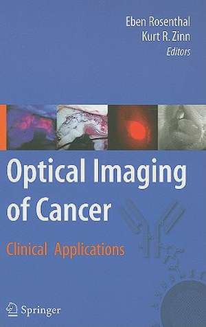
Optical Imaging of Cancer: Clinical Applications
Editat de Eben Rosenthal, Kurt R. Zinnen Limba Engleză Hardback – 12 oct 2009
Describe hardware adapted for small animal imaging and for clinical applications: endoscopes and operative microscopes.
Outline FDA approved and newer optical imaging probes. Include discussion of chemistry and linkage to other proteins. Review current techniques to image cancer and the development of techniques to specifically image cancer cells.
Review use of exploiting differences in tissue autofluorescence to diagnose and treat cancer. Include agents such as 5-aminoleculinic acid.
Review mechanisms that require proteolytic processing within the tumor to become active fluorophores.
Review use of cancer selective proteins to localize probes to cancer cells: include toxins, antibodies, and minibodies.
Introduction of plasmids, viruses or other genetic material may be used to express fluorescent agents in vivo. This chapter will review multiple vectors and delivery mechanisms of optical imaging cassettes.Preclinical investigations into the use of optical contrast agents for the detection of primary tumors in conventional and orthotopic models will be discussed.
Preclinical investigations into the use of optical contrast agents for the detection of metastatic tumors in mouse models will be discussed.
Use of targeted and non-specific optical contrast agents have been used for the detection of sentinel lymph node detection. These applications and how they differ from other applications will be discussed.
Because of the unique difficulty of identifying tumor from normal tissue in brain tissue, a separate chapter would be needed. More clinical data is available for this cancer type than any other.
Discussion of potential clinical applications for optical imaging and an assessment of the potential market.
| Toate formatele și edițiile | Preț | Express |
|---|---|---|
| Paperback (1) | 978.66 lei 38-44 zile | |
| Springer – 22 aug 2016 | 978.66 lei 38-44 zile | |
| Hardback (1) | 1103.03 lei 22-36 zile | |
| Springer – 12 oct 2009 | 1103.03 lei 22-36 zile |
Preț: 1103.03 lei
Preț vechi: 1161.08 lei
-5% Nou
Puncte Express: 1655
Preț estimativ în valută:
211.06€ • 220.96$ • 174.64£
211.06€ • 220.96$ • 174.64£
Carte disponibilă
Livrare economică 17-31 martie
Preluare comenzi: 021 569.72.76
Specificații
ISBN-13: 9780387938738
ISBN-10: 0387938737
Pagini: 272
Ilustrații: XIII, 272 p.
Dimensiuni: 155 x 235 x 18 mm
Greutate: 0.64 kg
Ediția:2010
Editura: Springer
Colecția Springer
Locul publicării:New York, NY, United States
ISBN-10: 0387938737
Pagini: 272
Ilustrații: XIII, 272 p.
Dimensiuni: 155 x 235 x 18 mm
Greutate: 0.64 kg
Ediția:2010
Editura: Springer
Colecția Springer
Locul publicării:New York, NY, United States
Public țintă
ResearchCuprins
Part I: Optical Imaging Principles Basic principles Kurt Zinn, University of Alabama at Birmingham Describe principles of optical imaging including chemistry and physics of fluorescence, limitations/advantages of optical imaging compared to metabolic and anatomic imaging. Hardware for imaging John Frangioni, Harvard University Describe hardware adapted for small animal imaging and for clinical applications: endoscopes and operative microscopes. Optical probes GE Healthcare Outline FDA approved and newer optical imaging probes. Include discussion of chemistry and linkage to other proteins. Part II: Cancer Targeting Strategies Introduction to molecular targeting strategies KE Adams, Baylor University Review current techniques to image cancer and the development of techniques to specifically image cancer cells. Tissue auto fluorescence CF Poh, Vancouver, Canada Review use of exploiting differences in tissue autofluorescence to diagnose and treat cancer. Include agents such as 5-aminoleculinic acid. Proteolytic mechanisms O. McIntyre, Vanderbilt University Review mechanisms that require proteolytic processing within the tumor to become active fluorophores. Antigen targeting M Garfinkle, Texas A&M University Review use of cancer selective proteins to localize probes to cancer cells: include toxins, antibodies, and minibodies. Genetically engineered optical imaging TBA Introduction of plasmids, viruses or other genetic material may be used to express fluorescent agents in vivo. This chapter will review multiple vectors and delivery mechanisms of optical imaging cassettes. Part III: Potential Clinical Applications Preclinical investigations: Detection of primary tumors Eben Rosenthal, UAB Preclinical investigations into the use of optical contrast agents for the detection of primary tumors in conventional and orthotopic models will be discussed. Preclinical investigations: Detection of metastatic cancer Y Hama, NIH/NCI Preclinical investigations into the use of optical contrast agents for the detection of metastatic tumors in mouse models will be discussed. Sentinel lymph node imaging P Wunderbaldinger, University of Vienna Use of targeted and non-specific optical contrast agents have been used for the detection of sentinel lymph node detection. These applications and how they differ from other applications will be discussed Neuro-oncologic applications Veiseh M, University of Seattle Because of the unique difficulty of identifying tumor from normal tissue in brain tissue, a separate chapter would be needed. More clinical data is available for this cancer type than any other. Future Directions for Clinical Use of Optical Imaging TBA Discussion of potential clinical applications for optical imaging and an assessment of the potential market.
Textul de pe ultima copertă
Optical detection represents the next great horizon for cancer imaging. As molecularly targeted therapeutic agents are delivered to the clinic in increasing numbers, there is a parallel opportunity to advance optical imaging techniques. Because of its limited toxicity and potential for real-time information, optical imaging represents an underdeveloped modality in clinical medicine. Optical Imaging of Cancer: Clinical Applications explores the preclinical and clinical data to support the use of these techniques in cancer imaging.
Different fluorescent delivery modalities are explored: from monoclonal antibodies to viral vector delivery. Indications for use include monitoring therapuetic delivery, early detection, and operative removal techniques in all stages of clinical development. This book represents the best and most clinically relevant techniques currently available.
Different fluorescent delivery modalities are explored: from monoclonal antibodies to viral vector delivery. Indications for use include monitoring therapuetic delivery, early detection, and operative removal techniques in all stages of clinical development. This book represents the best and most clinically relevant techniques currently available.
Caracteristici
Covers a wide array of areas including optical imaging principles and potential clinical applications
Includes supplementary material: sn.pub/extras
Includes supplementary material: sn.pub/extras
