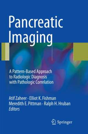
Pancreatic Imaging: A Pattern-Based Approach to Radiologic Diagnosis with Pathologic Correlation
Editat de Atif Zaheer, Elliot K. Fishman, Meredith E. Pittman, Ralph H. Hrubanen Limba Engleză Paperback – 12 aug 2018
| Toate formatele și edițiile | Preț | Express |
|---|---|---|
| Paperback (1) | 1156.07 lei 38-44 zile | |
| Springer International Publishing – 12 aug 2018 | 1156.07 lei 38-44 zile | |
| Hardback (1) | 1422.83 lei 38-44 zile | |
| Springer International Publishing – 16 aug 2017 | 1422.83 lei 38-44 zile |
Preț: 1156.07 lei
Preț vechi: 1216.92 lei
-5% Nou
Puncte Express: 1734
Preț estimativ în valută:
221.24€ • 230.13$ • 182.65£
221.24€ • 230.13$ • 182.65£
Carte tipărită la comandă
Livrare economică 08-14 aprilie
Preluare comenzi: 021 569.72.76
Specificații
ISBN-13: 9783319849614
ISBN-10: 3319849611
Pagini: 449
Ilustrații: XV, 449 p. 210 illus., 125 illus. in color.
Dimensiuni: 155 x 235 mm
Ediția:Softcover reprint of the original 1st ed. 2017
Editura: Springer International Publishing
Colecția Springer
Locul publicării:Cham, Switzerland
ISBN-10: 3319849611
Pagini: 449
Ilustrații: XV, 449 p. 210 illus., 125 illus. in color.
Dimensiuni: 155 x 235 mm
Ediția:Softcover reprint of the original 1st ed. 2017
Editura: Springer International Publishing
Colecția Springer
Locul publicării:Cham, Switzerland
Cuprins
Parenchymal abnormalities: Focal hypodense mass.- Diffuse Pancreatic hypodensity.- Cystic mass.- Solid hyperenhancing mass.- Gastrinoma.- Diffuse enlargement.- Ductal abnormalities.- Diffuse abnormalities.- Focal abnormalities.
Recenzii
“I really enjoyed reading through this book, with its numerous cases succinctly highlighting the essential imaging findings … . This would be a very useful text for the trainee and practising radiologist, and there are lots of interesting cases for the specialist. ... The case-based format with very good images, pathological correlation and excellent but concise discussion makes this a highly recommended book on pancreatic imaging.” (Dr Zahir Amin, RAD Magazine, April 2018)
Notă biografică
Atif Zaheer, MD, Johns Hopkins University, The Russell H. Morgan Department of Radiology and Radiological Science, Baltimore, MD, USA
Elliot K. Fishman, MD, Johns Hopkins University, The Russell H. Morgan Department of Radiology and Radiological Science, Baltimore, MD, USA
Ralph H. Hruban, MD, Johns Hopkins University, The Russell H. Morgan Department of Radiology and Radiological Science, Baltimore, MD, USA
Meredith Pittmann, MD, Johns Hopkins University, The Russell H. Morgan Department of Radiology and Radiological Science, Baltimore, MD, USA
Ralph H. Hruban, MD, Johns Hopkins University, The Russell H. Morgan Department of Radiology and Radiological Science, Baltimore, MD, USA
Meredith Pittmann, MD, Johns Hopkins University, The Russell H. Morgan Department of Radiology and Radiological Science, Baltimore, MD, USA
Textul de pe ultima copertă
This comprehensive teaching atlas covers virtually all pancreatic anatomy (including variants) and diseases in a pattern-based radiologic approach. Cases are presented as “unknowns”, allowing the reader to analyze the findings and learn key points. Each teaching case includes a brief clinical history, images, a description of imaging findings, differential diagnoses, final diagnosis with images of gross pathology, and a discussion of key teaching points. The presented images have been acquired with the full range of relevant modalities, including state of the art technologies such as multidetector row dual-phase CT, 3D reformatting, and multiple MRI sequences. The book will help radiologists, radiology residents and fellows to sharpen their diagnostic skills by looking at a vast array of pathology from a major tertiary hospital (Johns Hopkins) and will also assist in preparation for radiology board examinations.
Caracteristici
Covers virtually all anatomic variants and diseases in a pattern-based radiologic approach Offers informative correlation with histopathology Highlights important points relevant to everyday practice that all radiologists and pathologists need to know
