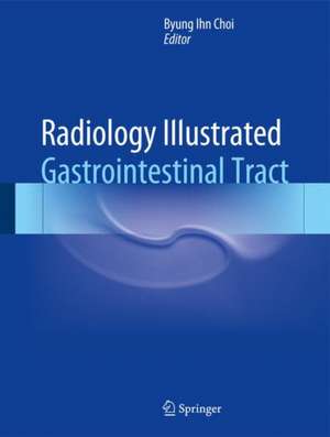
Radiology Illustrated: Gastrointestinal Tract: Radiology Illustrated
Editat de Byung Ihn Choien Limba Engleză Hardback – 29 sep 2014
| Toate formatele și edițiile | Preț | Express |
|---|---|---|
| Paperback (1) | 1673.01 lei 38-44 zile | |
| Springer Berlin, Heidelberg – oct 2016 | 1673.01 lei 38-44 zile | |
| Hardback (1) | 1847.64 lei 3-5 săpt. | |
| Springer Berlin, Heidelberg – 29 sep 2014 | 1847.64 lei 3-5 săpt. |
Preț: 1847.64 lei
Preț vechi: 1944.88 lei
-5% Nou
Puncte Express: 2771
Preț estimativ în valută:
353.59€ • 383.94$ • 297.01£
353.59€ • 383.94$ • 297.01£
Carte disponibilă
Livrare economică 01-15 aprilie
Preluare comenzi: 021 569.72.76
Specificații
ISBN-13: 9783642554117
ISBN-10: 3642554113
Pagini: 602
Ilustrații: XII, 602 p. 405 illus., 209 illus. in color.
Dimensiuni: 210 x 279 x 32 mm
Greutate: 1.89 kg
Ediția:2015
Editura: Springer Berlin, Heidelberg
Colecția Springer
Seria Radiology Illustrated
Locul publicării:Berlin, Heidelberg, Germany
ISBN-10: 3642554113
Pagini: 602
Ilustrații: XII, 602 p. 405 illus., 209 illus. in color.
Dimensiuni: 210 x 279 x 32 mm
Greutate: 1.89 kg
Ediția:2015
Editura: Springer Berlin, Heidelberg
Colecția Springer
Seria Radiology Illustrated
Locul publicării:Berlin, Heidelberg, Germany
Public țintă
Professional/practitionerCuprins
Part 1. Pharynx/Esophagus.- 1. Benign structural and functional abnormality of the esophagus, postop. Complication of the esophagus.- 2. Esophagitis.- 3. Benign and malignant tumor of the esophagus.- Part 2. Stomach/Duodenum.- 4. Benign structural & functional abnormality of the stomach and duodenum.- 5. Inflammatory and infectious diseases of the stomach and duodenum.- 6. Mucosal tumor of the stomach.- 7. Subepithelial tumor of the stomach.- 8. Lymphoma and metastasis of the stomach.- 9. Postoperative change and complications of the stomach and duodenum.- Part 3. Small Bowel.- 10. Benign structural & functional abnormality of the small bowel.- 11. Inflammatory disease of the small bowel.- 12. Vasculitis and ischemic bowel disease.- 13. Small bowel obstruction.- 14. Benign tumor of the small bowel.- 15. Malignant tumor of the small bowel.- Part 4. Colon.- 16. Benign structural & functional abnormality of the colon.- 17. Inflammatory & infectious disease of the colon.- 18. Benign tumor of the colon.- 19. Malignant tumor of the colon.- 20. Postoperative change and complications of the colon.- Part 5. Mesentery, Peritoneum, abdominal wall.- 21. Benign diseases of mesentery and omentum.- 22. Malignant diseases of mesentery and omentum.- 23. Abdominal wall abnormality & hernia.
Recenzii
“This book on gastrointestinal imaging has excellent images and a good variety of case collections. … The intended audience is radiology residents and general diagnostic radiology practitioners who have an interest in GI radiology. … This book definitely has a role as an excellent reference for board preparation as well as a guide to keep in a GI radiologist's personal library. … Its excellent endoscopic and pathology images distinguish it from other, similar books.” (Jayanth H. Keshavamurthy, Doody’s Book Reviews, September, 2015)
“This is a book on the radiology of the GI tract, including the mesentery, peritoneum and the abdominal wall. … this is overall an excellent book, with concise but comprehensive text followed by very good illustrations including histopathology and endoscopy images, which are an essential part of GI radiology. It is aimed at radiology trainees and established radiologists, as well as clinicians with an interest in GI imaging.” (Zahir Amin, RAD Magazine, July, 2015)
“This is a book on the radiology of the GI tract, including the mesentery, peritoneum and the abdominal wall. … this is overall an excellent book, with concise but comprehensive text followed by very good illustrations including histopathology and endoscopy images, which are an essential part of GI radiology. It is aimed at radiology trainees and established radiologists, as well as clinicians with an interest in GI imaging.” (Zahir Amin, RAD Magazine, July, 2015)
Notă biografică
Byung Ihn Choi MD, Seoul National University Hospital, Department of Radiology, 101 Daehak-ro, Jongno-gu, 110- 744 Seoul, Korea, Republic of (South Korea).
Textul de pe ultima copertă
Radiology Illustrated: Gastrointestinal Tract is the second of two volumes designed to provide clear and practical guidance on the diagnostic imaging of abdominal diseases. The book presents approximately 300 cases with 1500 carefully selected and categorized illustrations of gastrointestinal tract diseases, along with key text messages and tables that will help the reader easily to recall the relevant images as an aid to differential diagnosis., Essential points are summarized at the end of each text message to facilitate rapid review and learning. Additionally, brief descriptions of each clinical problem are provided, followed by case studies of both common and uncommon pathologies that illustrate the roles of the different imaging modalities, including ultrasound, radiography, computed tomography, and magnetic resonance imaging.
Caracteristici
Clear and practical guide to diagnostic imaging of diseases of the gastrointestinal tract Explains the roles of the different imaging modalities Aids differential diagnosis Includes a wealth of carefully selected and categorized illustrations Presents case studies of both common and uncommon pathologies
