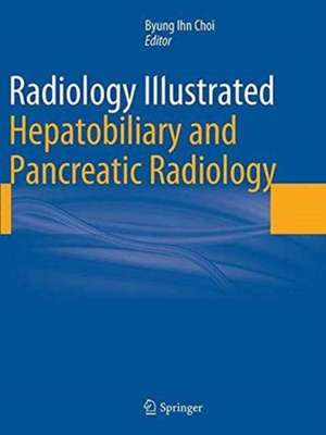
Radiology Illustrated: Hepatobiliary and Pancreatic Radiology: Radiology Illustrated
Editat de Byung Ihn Choien Limba Engleză Paperback – oct 2016
The book presents approximately 560 cases with more than 2100 carefully selected and categorized illustrations, along with key text messages and tables, that will allow the reader easily to recall the relevant images as an aid to differential diagnosis. At the end of each text message, key points are summarized to facilitate rapid review and learning. In addition, brief descriptions of each clinical problem are provided, followed by both common and uncommon case studies that illustrate the role of different imaging modalities, such as ultrasound, radiography, CT, and MRI.
| Toate formatele și edițiile | Preț | Express |
|---|---|---|
| Paperback (1) | 1673.01 lei 38-44 zile | |
| Springer Berlin, Heidelberg – oct 2016 | 1673.01 lei 38-44 zile | |
| Hardback (1) | 1847.64 lei 3-5 săpt. | |
| Springer Berlin, Heidelberg – 29 sep 2014 | 1847.64 lei 3-5 săpt. |
Preț: 1673.01 lei
Preț vechi: 1761.06 lei
-5% Nou
Puncte Express: 2510
Preț estimativ în valută:
320.23€ • 347.96$ • 269.17£
320.23€ • 347.96$ • 269.17£
Carte tipărită la comandă
Livrare economică 17-23 aprilie
Preluare comenzi: 021 569.72.76
Specificații
ISBN-13: 9783662523506
ISBN-10: 3662523507
Pagini: 852
Ilustrații: XVIII, 834 p. 585 illus., 185 illus. in color.
Dimensiuni: 210 x 279 x 47 mm
Ediția:Softcover reprint of the original 1st ed. 2014
Editura: Springer Berlin, Heidelberg
Colecția Springer
Seria Radiology Illustrated
Locul publicării:Berlin, Heidelberg, Germany
Cuprins
Part 1. Liver.- Anomalies and Anatomic Variants of the Liver.- Diffuse Liver Disease.- Benign Tumors of the Liver.- Hepatocellular Carcinoma.- Other Malignant Tumors of the Liver.- Focal Hepatic Infections.- Hemodynamic and Perfusion Related Disorders.- Liver Transplantation.- Therapeutic Response Evaluation of HCC.- Trauma and Post-treatment Complications of the Liver.- Part 2. Biliary tract.- Anomalies and Anatomic Variants of the Biliary tract.- Cholangitis.- Cholecystitis.- Cholangiocarcinoma.- Tumors of the Gallbladder.- Trauma and Post-treatment Complications of the Biliary tract.- Part 3. Pancreas.- Anomalies and Anatomic Variants of the Pancreas.- Pancreatitis.- Cystic Tumors of the Pancreas.- Solid Tumors of the Pancreas.- Trauma and Post-treatment Complications of the Pancreas.- Part 4. Spleen.- Anomalies and Anatomic Variants of the spleen.- Diffuse Spleen Disease.- Benign Focal Lesions of the Spleen.- Malignant Focal Lesions of the Spleen.- Trauma and Post-treatment Complications of the spleen.
Notă biografică
Byung Ihn Choi, MD, Ph.D. Department of Radiology, Seoul National University Hospital Professor, Seoul National University College of Medicine
Textul de pe ultima copertă
Radiology Illustrated: Hepatobiliary and Pancreatic Radiology is the first of two volumes that will serve as a clear, practical guide to the diagnostic imaging of abdominal diseases. This volume, devoted to diseases of the liver, biliary tree, gallbladder, pancreas, and spleen, covers congenital disorders, vascular diseases, benign and malignant tumors, and infectious conditions. Liver transplantation, evaluation of the therapeutic response of hepatocellular carcinoma, trauma, and post-treatment complications are also addressed.
The book presents approximately 560 cases with more than 2100 carefully selected and categorized illustrations, along with key text messages and tables, that will allow the reader easily to recall the relevant images as an aid to differential diagnosis. At the end of each text message, key points are summarized to facilitate rapid review and learning. In addition, brief descriptions of each clinical problem are provided, followed by both common and uncommon case studies that illustrate the role of different imaging modalities, such as ultrasound, radiography, CT, and MRI.
The book presents approximately 560 cases with more than 2100 carefully selected and categorized illustrations, along with key text messages and tables, that will allow the reader easily to recall the relevant images as an aid to differential diagnosis. At the end of each text message, key points are summarized to facilitate rapid review and learning. In addition, brief descriptions of each clinical problem are provided, followed by both common and uncommon case studies that illustrate the role of different imaging modalities, such as ultrasound, radiography, CT, and MRI.
Caracteristici
Clear, practical guide to the diagnostic imaging of diseases of the liver, biliary tree, gallbladder, pancreas, and spleen A wealth of carefully selected and categorized illustrations Highlighted key points to facilitate rapid review Aid to differential diagnosis
Recenzii
“This book on gastrointestinal imaging has excellent images and a good variety of case collections. … The intended audience is radiology residents and general diagnostic radiology practitioners who have an interest in GI radiology. … This book definitely has a role as an excellent reference for board preparation as well as a guide to keep in a GI radiologist's personal library. … Its excellent endoscopic and pathology images distinguish it from other, similar books.” (Jayanth H. Keshavamurthy, Doody’s Book Reviews, September, 2015)
“This is a book on the radiology of the GI tract, including the mesentery, peritoneum and the abdominal wall. … this is overall an excellent book, with concise but comprehensive text followed by very good illustrations including histopathology and endoscopy images, which are an essential part of GI radiology. It is aimed at radiology trainees and established radiologists, as well as clinicians with an interest in GI imaging.” (Zahir Amin, RAD Magazine, July, 2015)
“This is a book on the radiology of the GI tract, including the mesentery, peritoneum and the abdominal wall. … this is overall an excellent book, with concise but comprehensive text followed by very good illustrations including histopathology and endoscopy images, which are an essential part of GI radiology. It is aimed at radiology trainees and established radiologists, as well as clinicians with an interest in GI imaging.” (Zahir Amin, RAD Magazine, July, 2015)
