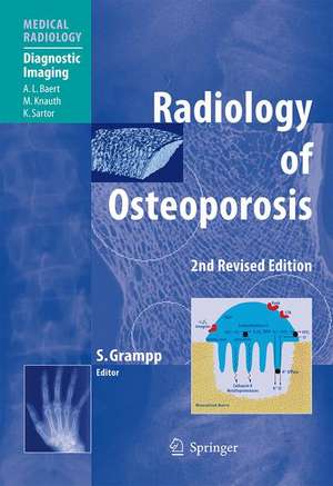
Radiology of Osteoporosis: Medical Radiology
Editat de Stephan Grampp Cuvânt înainte de A.L. Baerten Limba Engleză Hardback – 22 feb 2008
| Toate formatele și edițiile | Preț | Express |
|---|---|---|
| Paperback (1) | 719.29 lei 38-44 zile | |
| Springer Berlin, Heidelberg – 12 feb 2010 | 719.29 lei 38-44 zile | |
| Hardback (1) | 989.22 lei 38-44 zile | |
| Springer Berlin, Heidelberg – 22 feb 2008 | 989.22 lei 38-44 zile |
Din seria Medical Radiology
- 5%
 Preț: 1108.87 lei
 - 5%
 Preț: 349.24 lei
 - 5%
 Preț: 1308.02 lei
 - 5%
 Preț: 1308.74 lei
 - 5%
 Preț: 720.68 lei
 - 5%
 Preț: 717.20 lei
 - 5%
 Preț: 1626.03 lei
 - 5%
 Preț: 1618.70 lei
 - 5%
 Preț: 802.21 lei
 - 5%
 Preț: 1130.07 lei
 - 5%
 Preț: 1116.00 lei
 - 5%
 Preț: 1953.34 lei
 - 5%
 Preț: 783.04 lei
 - 5%
 Preț: 1105.61 lei
 - 5%
 Preț: 794.00 lei
 - 5%
 Preț: 1101.21 lei
 - 5%
 Preț: 821.19 lei
 - 5%
 Preț: 1420.29 lei
 - 5%
 Preț: 743.16 lei
 - 5%
 Preț: 906.63 lei
 - 5%
 Preț: 1313.75 lei
 - 5%
 Preț: 1858.30 lei
 - 5%
 Preț: 1306.73 lei
 - 5%
 Preț: 1113.11 lei
 - 5%
 Preț: 1462.37 lei
 - 5%
 Preț: 1301.44 lei
 - 5%
 Preț: 975.17 lei
 - 5%
 Preț: 1122.58 lei
 - 5%
 Preț: 1986.27 lei
 - 5%
 Preț: 1126.82 lei
 - 5%
 Preț: 718.46 lei
 - 5%
 Preț: 1450.84 lei
 - 5%
 Preț: 1298.14 lei
 - 5%
 Preț: 1110.32 lei
 - 5%
 Preț: 1184.42 lei
 - 5%
 Preț: 1113.99 lei
 - 5%
 Preț: 1435.85 lei
 - 5%
 Preț: 663.23 lei
 - 5%
 Preț: 1605.11 lei
 - 5%
 Preț: 731.07 lei
 - 5%
 Preț: 733.09 lei
 - 5%
 Preț: 1124.07 lei
 - 5%
 Preț: 383.93 lei
 - 5%
 Preț: 1106.69 lei
 - 5%
 Preț: 982.50 lei
 - 5%
 Preț: 1317.17 lei
 - 5%
 Preț: 1437.67 lei
 - 5%
 Preț: 1307.85 lei
 - 5%
 Preț: 1950.60 lei
Preț: 989.22 lei
Preț vechi: 1041.27 lei
-5% Nou
Puncte Express: 1484
Preț estimativ în valută:
189.31€ • 205.56$ • 159.02£
189.31€ • 205.56$ • 159.02£
Carte tipărită la comandă
Livrare economică 19-25 aprilie
Preluare comenzi: 021 569.72.76
Specificații
ISBN-13: 9783540258889
ISBN-10: 3540258884
Pagini: 260
Ilustrații: X, 247 p.
Dimensiuni: 193 x 270 x 20 mm
Greutate: 0.87 kg
Ediția:2nd rev. ed. 2008
Editura: Springer Berlin, Heidelberg
Colecția Springer
Seriile Medical Radiology, Diagnostic Imaging
Locul publicării:Berlin, Heidelberg, Germany
ISBN-10: 3540258884
Pagini: 260
Ilustrații: X, 247 p.
Dimensiuni: 193 x 270 x 20 mm
Greutate: 0.87 kg
Ediția:2nd rev. ed. 2008
Editura: Springer Berlin, Heidelberg
Colecția Springer
Seriile Medical Radiology, Diagnostic Imaging
Locul publicării:Berlin, Heidelberg, Germany
Public țintă
Professional/practitionerCuprins
to Bone Development, Remodelling and Repair.- Pathophysiology and Aging of Bone.- Pathophysiology of Rheumatoid Arthritis and Other Disorders.- Therapeutic Approaches and Mechanisms of Drug Action.- Orthopedic Surgery.- Radiology of Osteoporosis.- Dual-Energy X-Ray Absorptiometry.- Vertebral Morphometry.- Spinal Quantitative Computed Tomography.- pQCT: Peripheral Quantitative Computed Tomography.- Quantitative Ultrasound.- Magnetic Resonance Imaging.- Structure Analysis Using High-Resolution Imaging Techniques.- Densitometry in Clinical Practice.- Practical Cases.
Recenzii
From the reviews of the second edition:
"‘Radiology of Osteoporosis,’ … is a revised edition of a volume in the Springer Medical Radiology series first published in 2002. The book is intended to provide a guide for radiologists, orthopaedic surgeons and other specialists on osteoporosis and the various medical imaging techniques that assist in its diagnosis. … This book gives a useful and reasonably up-to-date account of the basic measurement techniques used in bone densitometry." (Glen Blake, RAD magazine, January, 2009)
"It provides a comprehensive overview of the different aspects of osteoporosis including the pathologic conditions that give rise to osteoporosis and the complications that frequently occur. Well-known international authors provide their most current data on morphology, pathophysiology and therapeutic approaches in osteoporosis … . In summary, the information provided in this book will be invaluable to radiologists and all clinicians involved in the care of patients with osteoporosis." (A. K. Dixon and Thomas J. Vogl, European Radiology, Vol. 19, 2009)
Textul de pe ultima copertă
This second edition of Radiology of Osteoporosis has been fully updated so as to represent the current state of the art. It provides a comprehensive overview of osteoporosis, the pathologic conditions that give rise to osteoporosis, and the complications that are frequently encountered. After initial chapters devoted to pathophysiology, the presentation of osteoporosis on conventional radiographs is illustrated and discussed. Thereafter, detailed consideration is given to each of the measurement methods employed to evaluate osteoporosis, including dual x-ray absorptiometry, vertebral morphometry, spinal and peripheral quantitative computed tomography, quantitative ultrasound, and magnetic resonance imaging. The role of densitometry in daily clinical practice is appraised. Finally, a collection of difficult cases involving pitfalls is presented, with guidance to their solution. The information contained in this volume will be invaluable to all with an interest in osteoporosis.
Caracteristici
Documents the radiographic presentation of osteoporosis Focuses on the reasons for particular imaging findings Discusses in detail the various quantitative measurement methods and their use Illustrates pitfalls and offers guidance on avoiding misinterpretation Completely updated to represent the current state of the art Includes supplementary material: sn.pub/extras
