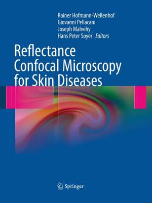
Reflectance Confocal Microscopy for Skin Diseases
Editat de Rainer Hofmann-Wellenhof, Giovanni Pellacani, Joseph Malvehy, H. Peter Soyeren Limba Engleză Paperback – 23 aug 2016
| Toate formatele și edițiile | Preț | Express |
|---|---|---|
| Paperback (1) | 1123.33 lei 6-8 săpt. | |
| Springer Berlin, Heidelberg – 23 aug 2016 | 1123.33 lei 6-8 săpt. | |
| Hardback (1) | 1646.52 lei 3-5 săpt. | |
| Springer Berlin, Heidelberg – mar 2012 | 1646.52 lei 3-5 săpt. |
Preț: 1123.33 lei
Preț vechi: 1182.46 lei
-5% Nou
Puncte Express: 1685
Preț estimativ în valută:
215.02€ • 233.64$ • 180.73£
215.02€ • 233.64$ • 180.73£
Carte tipărită la comandă
Livrare economică 21 aprilie-05 mai
Preluare comenzi: 021 569.72.76
Specificații
ISBN-13: 9783662501887
ISBN-10: 3662501880
Pagini: 500
Ilustrații: IX, 500 p.
Dimensiuni: 210 x 279 mm
Greutate: 1.14 kg
Ediția:Softcover reprint of the original 1st ed. 2012
Editura: Springer Berlin, Heidelberg
Colecția Springer
Locul publicării:Berlin, Heidelberg, Germany
ISBN-10: 3662501880
Pagini: 500
Ilustrații: IX, 500 p.
Dimensiuni: 210 x 279 mm
Greutate: 1.14 kg
Ediția:Softcover reprint of the original 1st ed. 2012
Editura: Springer Berlin, Heidelberg
Colecția Springer
Locul publicării:Berlin, Heidelberg, Germany
Cuprins
From the Contents: CONFOCAL REFLECTANCE MICROSCOPY – THE ESSENTIALS.- NORMAL SKIN.- MELANOCYTIC LESIONS.- MELANOCYTIC NEVI.- MELANOMA.- Superficial Spreading Melanoma.- Melanoma Progression.- Nodular Melanoma.- Lentigo Maligna.- Amelanotic Melanoma.- NON-MELANOCYTIC SKIN LESIONS.- Semeiology and Pattern Analysis In Non-Melanocytic Lesions.- Dermoscopic and Histologic Correlations.- Solar Lentigo, Seborrheic Keratosis and Lichen Planus Like-Keratosi.- Basal Cell Carcinoma.- Actinic Keratoses.- Squamous Cell Carcinoma .- Cutaneous Lymphoma.- Potpourri of Non Melanocytic Skin Lesions.- INFLAMMATORY SKIN DISEASES.- MONITORING OF SKIN LESIONS AND THERAPY CONTROL.- FUTURE ASPECTS.- Tele-Reflectance Confocal Microscopy.- Automated Diagnosis and Reflectance Confocal Microscopy.- Experimental Applications and Future Directions.- CONSENSUS TERMINOLOGY GLOSSARY.
Notă biografică
G. Pellacani, MD, is Professor of Dermatology in the Department of Dermatology, University of Modena and Reggio Emilia, Italy. His major research interest is in the field of melanoma diagnosis by means of non-invasive techniques such as dermoscopy and in vivo confocal microscopy.
Josep Malvehy, MD, is Director of the Melanoma Unit at the University Hospital Clinic of Barcelona, Spain. His principal interest lies in melanoma and skin cancer, including diagnosis with non-invasive imaging technology.
H. Peter Soyer, MD, is Chair of the Dermatology Research Centre at the University of Queensland in Brisbane, Australia. He has had a longstanding interest in cutaneous bio-optical imaging techniques and clinicopathologic correlation of cutaneous neoplasms.
Rainer Hofmann-Wellenhof, MD, is Professor of Dermatology in the Department of Dermatology, Medical University of Graz, Austria and Director of the Pigmented Skin Lesion Clinic at the Department of Dermatology in Graz. He has been a dermato-oncologist for 20 years. His special interests include diagnostic methods for skin tumors and preventive dermato-oncology.
Textul de pe ultima copertă
In recent years the relevance of non-invasive bioimaging techniques in the field of melanoma screening has steadily increased. In the new era of “clinicoimaging” diagnosis, reflectance confocal microscopy (RCM) will have a major impact on the diagnosis and management of neoplastic and inflammatory skin diseases.
This book focuses on the use and significance of in vivo RCM for non-invasive high-resolution imaging of the skin. All of the chapters in this hands-on guide are generously illustrated with numerous confocal images and structured in a reader-friendly way. The contents include:
• detailed information on the most relevant and up-to-date aspects of RCM,
• schematic drawings summarizing and explaining the most important RCM criteria, and
• a chapter specifically devoted to bridging the gap between dermoscopy, RCM, and histopathology.
At the end of each chapter, core messages recapitulate the most pertinent aspects.
Reflectance Confocal Microscopy for Skin Diseases will be a valuable resource for all physicians involved in the diagnosis and treatment of neoplastic and inflammatory skin diseases
Caracteristici
This unique book encompasses the most relevant and up-to-date aspects of RCM (reflectance confocal microscopy) It serves as a helpful hands-on guide for confocal imaging for dermatologists It includes a chapter devoted to bridging the gap between dermoscopy, RCM and histopathology All chapters are lavishly illustrated and reader-friendly structured
