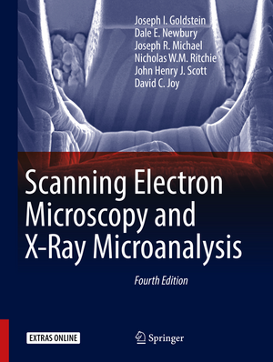
Scanning Electron Microscopy and X-Ray Microanalysis
Autor Joseph I. Goldstein, Dale E. Newbury, Joseph R. Michael, Nicholas W.M. Ritchie, John Henry J. Scott, David C. Joyen Limba Engleză Hardback – 18 noi 2017
- Realigns
the
text
with
the
needs
of
a
diverse
audience
from
researchers
and
graduate
students
to
SEM
operators
and
technical
managers
- Emphasizes
practical,
hands-on
operation
of
the
microscope,
particularly
user
selection
of
the
critical
operating
parameters
to
achieve
meaningful
results
- Provides
step-by-step
overviews
of
SEM,
EDS,
and
EBSD
and
checklists
of
critical
issues
for
SEM
imaging,
EDS
x-ray
microanalysis,
and
EBSD
crystallographic
measurements
- Makes
extensive
use
of
open
source
software:
NIH
ImageJ-FIJI
for
image
processing
and
NIST
DTSA
II
for
quantitative
EDS
x-ray
microanalysis
and
EDS
spectral
simulation.
- Includes
case
studies
to
illustrate
practical
problem
solving
- Covers
Helium
ion
scanning
microscopy
- Organized
into
relatively
self-contained
modules
–
no
need
to
"read
it
all"
to
understand
a
topic
- Includes an online supplement—an extensive "Database of Electron–Solid Interactions"—which can be accessed on SpringerLink, in Chapter 3
| Toate formatele și edițiile | Preț | Express |
|---|---|---|
| Paperback (1) | 621.41 lei 38-44 zile | |
| Springer – 30 aug 2018 | 621.41 lei 38-44 zile | |
| Hardback (1) | 643.53 lei 3-5 săpt. | +74.32 lei 7-13 zile |
| Springer – 18 noi 2017 | 643.53 lei 3-5 săpt. | +74.32 lei 7-13 zile |
Preț: 643.53 lei
Preț vechi: 804.42 lei
-20% Nou
123.14€ • 128.57$ • 101.91£
Carte disponibilă
Livrare economică 15-29 martie
Livrare express 01-07 martie pentru 84.31 lei
Specificații
ISBN-10: 149396674X
Pagini: 510
Ilustrații: XXIII, 550 p. 546 illus., 409 illus. in color. With online files/update.
Dimensiuni: 210 x 279 x 36 mm
Greutate: 2 kg
Ediția:4th ed. 2018
Editura: Springer
Colecția Springer
Locul publicării:New York, NY, United States
Cuprins
Preface.- Scanning Electron Microscopy and Associated Techniques: Overview.- Electron Beam – Specimen Interactions: Interaction Volume.- Backscattered Electrons.- Secondary Electrons.- X-rays.- SEM Instrumentation.- Image Formation.- SEM Image Interpretation.- The Visibility of Features in SEM Images.- Image Defects.- High resolution imaging.- Low Beam Energy SEM.- Variable Pressure Scanning Electron Microscopy (VPSEM).- ImageJ and Fiji.- SEM Imaging checklist.- SEM Case Studies.- Energy Dispersive X-ray Spectrometry: Physical Principles and User-Selected Parameters.- DTSA-II EDS Software.- Qualitative Elemental Analysis by Energy Dispersive X-ray Spectrometry.- Quantitative Analysis: from k-ratio to Composition.- Quantitative analysis: the SEM/EDS elemental microanalysis k-ratio procedure for bulk specimens, step-by-step.- Trace Analysis by SEM/EDS.- Low Beam Energy X-ray Microanalysis.- Analysis of Specimens with Special Geometry: Irregular Bulk Objects and Particles.- Compositional Mapping.- Attempting Electron-Excited X-ray Microanalysis in the Variable Pressure Scanning Electron Microscope (VP-SEM).- Energy Dispersive X-ray Microanalysis Checklist.- X-ray Microanalysis Case Studies.- Cathodoluminescence.- Characterizing crystalline materials in the SEM.- Focused Ion Beam Applications in the SEM laboratory.- Ion Beam Microscopy.- Appendix – A Database of Electron-Solid Interactions.- Index.
Recenzii
“There is no other single volume that covers as much theory and practice of SEM or X-ray microanalysis as Scanning Electron Microscopy and X-ray Microanalysis, 3rd Edition does. It is clearly written ... well organized. ... This is a reference text that no SEM or EPMA laboratory should be without.” (Thomas J. Wilson, Scanning, Vol. 27 (4), July/August, 2005)
“As the authors pointed out, the number of equations in the book is kept to a minimum, and important conceptions are also explained in a qualitative manner. A lot of very distinct images and schematic drawings make for a very interesting book and help readers who study scanning electron microscopy and X-ray microanalysis. The principal application and sample preparation given in this book are suitable for undergraduate students and technicians learning SEEM and EDS/WDS analyses. It is an excellent textbook for graduate students, and an outstanding reference for engineers, physical, and biological scientists.” (Microscopy and Microanalysis, Vol. 9 (5), October, 2003)
Notă biografică
This text is written by a team of authors associated with SEM and X-ray Microanalysis Courses presented as part of the Lehigh University Microscopy Summer School. Several of the authors have participated in this activity for more than 30 years.
Textul de pe ultima copertă
- Realigns the text with the needs of a diverse audience from researchers and graduate students to SEM operators and technical managers
- Emphasizes practical, hands-on operation of the microscope, particularly user selection of the critical operating parameters to achieve meaningful results
- Provides step-by-step overviews of SEM, EDS, and EBSD and checklists of critical issues for SEM imaging, EDS x-ray microanalysis, and EBSD crystallographic measurements
- Makes extensive use of open source software: NIH ImageJ-FIJI for image processing and NIST DTSA II for quantitative EDS x-ray microanalysis and EDS spectral simulation.
- Includes case studies to illustrate practical problem solving
- Covers Helium ion scanning microscopy
- Organized into relatively self-contained modules – no need to "read it all" to understand a topic
- Includes an online supplement—an extensive "Database of Electronic–Solid Interactions"—which can be accessed on SpringerLink, in Chapter 3
Caracteristici
Descriere
- Realigns the text with the needs of a diverse audience from researchers and graduate students to SEM operators and technical managers
- Emphasizes practical, hands-on operation of the microscope, particularly user selection of the critical operating parameters to achieve meaningful results
- Provides step-by-step overviews of SEM, EDS, and EBSD and checklists of critical issues for SEM imaging, EDS x-ray microanalysis, and EBSD crystallographic measurements
- Makes extensive use of open source software: NIH ImageJ-FIJI for image processing and NIST DTSA II for quantitative EDS x-ray microanalysis and EDS spectral simulation.
- Includes case studies to illustrate practical problem solving
- Covers Helium ion scanning microscopy
- Organized into relatively self-contained modules – no need to "read it all" to understand a topic
- Includes an online supplement—an extensive "Database of Electron–Solid Interactions"—which can be accessed on SpringerLink, in Chapter 3
