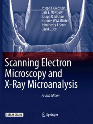
Scanning Electron Microscopy and X-Ray Microanalysis
Autor Joseph I. Goldstein, Dale E. Newbury, Joseph R. Michael, Nicholas W.M. Ritchie, John Henry J. Scott, David C. Joyen Limba Engleză Paperback – 30 aug 2018
This thoroughly revised and updated Fourth Edition of a time-honored text provides the reader with a comprehensive introduction to the field of scanning electron microscopy (SEM), energy dispersive X-ray spectrometry (EDS) for elemental microanalysis, electron backscatter diffraction analysis (EBSD) for micro-crystallography, and focused ion beams. Students and academic researchers will find the text to be an authoritative and scholarly resource, while SEM operators and a diversity of practitioners — engineers, technicians, physical and biological scientists, clinicians, and technical managers — will find that every chapter has been overhauled to meet the more practical needs of the technologist and working professional. In a break with the past, this Fourth Edition de-emphasizes the design and physical operating basis of the instrumentation, including the electron sources, lenses, detectors, etc. In the modern SEM, many of the low level instrument parameters are now controlled and optimized by the microscope’s software, and user access is restricted. Although the software control system provides efficient and reproducible microscopy and microanalysis, the user must understand the parameter space wherein choices are made to achieve effective and meaningful microscopy, microanalysis, and micro-crystallography. Therefore, special emphasis is placed on beam energy, beam current, electron detector characteristics and controls, and ancillary techniques such as energy dispersive x-ray spectrometry (EDS) and electron backscatter diffraction (EBSD).
With 13 years between the publication of the third and fourth editions, new coverage reflects the many improvements in the instrument and analysis techniques. The SEM has evolved into a powerful and versatile characterization platform in which morphology, elemental composition, and crystal structure can be evaluated simultaneously. Extension of the SEM into a "dual beam" platform incorporating bothelectron and ion columns allows precision modification of the specimen by focused ion beam milling. New coverage in the Fourth Edition includes the increasing use of field emission guns and SEM instruments with high resolution capabilities, variable pressure SEM operation, theory, and measurement of x-rays with high throughput silicon drift detector (SDD-EDS) x-ray spectrometers. In addition to powerful vendor- supplied software to support data collection and processing, the microscopist can access advanced capabilities available in free, open source software platforms, including the National Institutes of Health (NIH) ImageJ-Fiji for image processing and the National Institute of Standards and Technology (NIST) DTSA II for quantitative EDS x-ray microanalysis and spectral simulation, both of which are extensively used in this work. However, the user has a responsibility to bring intellect, curiosity, and a proper skepticism to information on a computer screen and to the entire measurement process. This book helps you to achieve this goal.
- Realigns the text with the needs of a diverse audience from researchers and graduate students to SEM operators and technical managers
- Emphasizes practical, hands-on operation of the microscope, particularly user selection of the critical operating parameters to achieve meaningful results
- Provides step-by-step overviews of SEM, EDS, and EBSD and checklists of critical issues for SEM imaging, EDS x-ray microanalysis, and EBSD crystallographic measurements
- Makes extensive use of open source software: NIH ImageJ-FIJI for image processing and NIST DTSA II for quantitative EDS x-ray microanalysis and EDS spectral simulation.
- Includes case studies to illustrate practical problem solving
- Covers Helium ion scanning microscopy
- Organized into relatively self-contained modules – no need to "read it all" to understand a topic
- Includesan online supplement—an extensive "Database of Electron–Solid Interactions"—which can be accessed on SpringerLink, in Chapter 3
| Toate formatele și edițiile | Preț | Express |
|---|---|---|
| Paperback (1) | 621.41 lei 38-44 zile | |
| Springer – 30 aug 2018 | 621.41 lei 38-44 zile | |
| Hardback (1) | 643.53 lei 3-5 săpt. | +74.32 lei 7-13 zile |
| Springer – 18 noi 2017 | 643.53 lei 3-5 săpt. | +74.32 lei 7-13 zile |
Preț: 621.41 lei
Preț vechi: 767.17 lei
-19% Nou
Puncte Express: 932
Preț estimativ în valută:
118.90€ • 124.15$ • 98.41£
118.90€ • 124.15$ • 98.41£
Carte tipărită la comandă
Livrare economică 01-07 aprilie
Preluare comenzi: 021 569.72.76
Specificații
ISBN-13: 9781493982691
ISBN-10: 1493982699
Pagini: 550
Ilustrații: XXIII, 550 p. 546 illus., 409 illus. in color.
Dimensiuni: 210 x 279 x 33 mm
Greutate: 1.5 kg
Ediția:4th ed. 2018
Editura: Springer
Colecția Springer
Locul publicării:New York, NY, United States
ISBN-10: 1493982699
Pagini: 550
Ilustrații: XXIII, 550 p. 546 illus., 409 illus. in color.
Dimensiuni: 210 x 279 x 33 mm
Greutate: 1.5 kg
Ediția:4th ed. 2018
Editura: Springer
Colecția Springer
Locul publicării:New York, NY, United States
Cuprins
Preface.- Scanning Electron Microscopy and Associated Techniques: Overview.- Electron Beam – Specimen Interactions: Interaction Volume.- Backscattered Electrons.- Secondary Electrons.- X-rays.- SEM Instrumentation.- Image Formation.- SEM Image Interpretation.- The Visibility of Features in SEM Images.- Image Defects.- High resolution imaging.- Low Beam Energy SEM.- Variable Pressure Scanning Electron Microscopy (VPSEM).- ImageJ and Fiji.- SEM Imaging checklist.- SEM Case Studies.- Energy Dispersive X-ray Spectrometry: Physical Principles and User-Selected Parameters.- DTSA-II EDS Software.- Qualitative Elemental Analysis by Energy Dispersive X-ray Spectrometry.- Quantitative Analysis: from k-ratio to Composition.- Quantitative analysis: the SEM/EDS elemental microanalysis k-ratio procedure for bulk specimens, step-by-step.- Trace Analysis by SEM/EDS.- Low Beam Energy X-ray Microanalysis.- Analysis of Specimens with Special Geometry: Irregular Bulk Objects and Particles.- CompositionalMapping.- Attempting Electron-Excited X-ray Microanalysis in the Variable Pressure Scanning Electron Microscope (VP-SEM).- Energy Dispersive X-ray Microanalysis Checklist.- X-ray Microanalysis Case Studies.- Cathodoluminescence.- Characterizing crystalline materials in the SEM.- Focused Ion Beam Applications in the SEM laboratory.- Ion Beam Microscopy.- Appendix – A Database of Electron-Solid Interactions.- Index.
Recenzii
Form the reviews of the third edition:
“There is no other single volume that covers as much theory and practice of SEM or X-ray microanalysis as Scanning Electron Microscopy and X-ray Microanalysis, 3rd Edition does. It is clearly written ... well organized. ... This is a reference text that no SEM or EPMA laboratory should be without.” (Thomas J. Wilson, Scanning, Vol. 27 (4), July/August, 2005)
“As the authors pointed out, the number of equations in the book is kept to a minimum, and important conceptions are also explained in a qualitative manner. A lot of very distinct images and schematic drawings make for a very interesting book and help readers who study scanning electron microscopy and X-ray microanalysis. The principal application and sample preparation given in this book are suitable for undergraduate students and technicians learning SEEM and EDS/WDS analyses. It is an excellent textbook for graduate students, and an outstanding reference for engineers, physical, and biological scientists.” (Microscopy and Microanalysis, Vol. 9 (5), October, 2003)
Notă biografică
This text is written by a team of authors associated with SEM and X-ray Microanalysis Courses presented as part of the Lehigh University Microscopy Summer School. Several of the authors have participated in this activity for more than 30 years.
Textul de pe ultima copertă
This thoroughly revised and updated Fourth Edition of a time-honored text provides the reader with a comprehensive introduction to the field of scanning electron microscopy (SEM), energy dispersive X-ray spectrometry (EDS) for elemental microanalysis, electron backscatter diffraction analysis (EBSD) for micro-crystallography and focused ion beams. Students and academic researchers will find the text to be an authoritative and scholarly resource, while SEM operators and a diversity of practitioners — engineers, technicians, physical and biological scientists, clinicians, and technical managers — will find that every chapter has been overhauled to meet the more practical needs of the technologist and working professional. In a break with the past, this Fourth Edition de-emphasizes the design and physical operating basis of the instrumentation, including the electron sources, lenses, detectors, etc. In the modern SEM, many of the low level instrument parameters are now controlled andoptimized by the microscope’s software, and user access is restricted. Although the software control system provides efficient and reproducible microscopy and microanalysis, the user must understand the parameter space wherein choices are made to achieve effective andmeaningful microscopy, microanalysis, and micro-crystallography. Therefore, special emphasis is placed on beam energy, beam current, electron detector characteristics and controls, and ancillary techniques such as energy dispersive x-ray spectrometry (EDS) and electron backscatter diffraction (EBSD).
With 13 years between the publication of the third and fourth editions, new coverage reflects the many improvements in the instrument and analysis techniques. The SEM has evolved into a powerful and versatile characterization platform in which morphology, elemental composition, and crystal structure can be evaluated simultaneously. Extension of the SEM into a "dual beam" platform incorporating both electron and ion columns allows precision modification of the specimen by focused ion beam milling. New coverage in the Fourth Edition includes the increasing use of field emission guns and SEM instruments with high resolution capabilities, variable pressure SEM operation, theory, and measurement of x-rays with high throughput silicon drift detector (SDD-EDS) x-ray spectrometers. In addition to powerful vendor- supplied software to support data collection and processing, the microscopist can access advanced capabilities available in free, open source software platforms, including the National Institutes of Health (NIH) ImageJ-Fiji for image processing and the National Institute of Standards and Technology (NIST) DTSA II for quantitative EDS x-ray microanalysis and spectral simulation, both of which are extensively used in this work. However, the user has a responsibility to bring intellect, curiosity, and a proper skepticism to information on a computer screen and to the entire measurement process. This book helps you to achieve this goal.
- Realigns the text with the needs of a diverse audience from researchers and graduate students to SEM operators and technical managers
- Emphasizes practical, hands-on operation of the microscope, particularly user selection of the critical operating parameters to achieve meaningful results
- Provides step-by-step overviews of SEM, EDS, and EBSD and checklists of critical issues for SEM imaging, EDS x-ray microanalysis, and EBSD crystallographic measurements
- Makes extensive use of open source software: NIH ImageJ-FIJI for image processing and NIST DTSA II for quantitative EDS x-ray microanalysis and EDS spectral simulation.
- Includes case studies to illustrate practical problem solving
- Covers Helium ion scanning microscopy
- Organized into relatively self-contained modules – no need to "read it all" to understand a topic
- Includes an online supplement—an extensive "Database of Electronic–Solid Interactions"—which can be accessed on SpringerLink, in Chapter 3
Caracteristici
Realigns the text with the needs of a diverse audience from researchers and graduate students to SEM operators and technical managers Emphasizes practical, hands-on operation of the microscope, particularly user selection of the critical operating parameters to achieve meaningful results Provides step-by-step overviews of SEM, EDS, and EBSD and checklists of critical issues for SEM imaging, EDS x-ray microanalysis, and EBSD crystallographic measurements Makes extensive use of open source software: NIH ImageJ-FIJI for image processing and NIST DTSA II for quantitative EDS x-ray microanalysis and EDS spectral simulation Includes case studies to illustrate practical problem solving Covers Helium ion scanning microscopy Organized into relatively self-contained modules – no need to "read it all" to understand a topic
Descriere
Descriere de la o altă ediție sau format:
This thoroughly revised and updated Fourth Edition of a time-honored text provides the reader with a comprehensive introduction to the field of scanning electron microscopy (SEM), energy dispersive X-ray spectrometry (EDS) for elemental microanalysis, electron backscatter diffraction analysis (EBSD) for micro-crystallography, and focused ion beams. Students and academic researchers will find the text to be an authoritative and scholarly resource, while SEM operators and a diversity of practitioners — engineers, technicians, physical and biological scientists, clinicians, and technical managers — will find that every chapter has been overhauled to meet the more practical needs of the technologist and working professional. In a break with the past, this Fourth Edition de-emphasizes the design and physical operating basis of the instrumentation, including the electron sources, lenses, detectors, etc. In the modern SEM, many of the low level instrument parameters are now controlled and optimized by the microscope’s software, and user access is restricted. Although the software control system provides efficient and reproducible microscopy and microanalysis, the user must understand the parameter space wherein choices are made to achieve effective and meaningful microscopy, microanalysis, and micro-crystallography. Therefore, special emphasis is placed on beam energy, beam current, electron detector characteristics and controls, and ancillary techniques such as energy dispersive x-ray spectrometry (EDS) and electron backscatter diffraction (EBSD).
With 13 years between the publication of the third and fourth editions, new coverage reflects the many improvements in the instrument and analysis techniques. The SEM has evolved into a powerful and versatile characterization platform in which morphology, elemental composition, and crystal structure can be evaluated simultaneously. Extension of the SEM into a "dual beam" platform incorporating both electron and ion columns allows precision modification of the specimen by focused ion beam milling. New coverage in the Fourth Edition includes the increasing use of field emission guns and SEM instruments with high resolution capabilities, variable pressure SEM operation, theory, and measurement of x-rays with high throughput silicon drift detector (SDD-EDS) x-ray spectrometers. In addition to powerful vendor- supplied software to support data collection and processing, the microscopist can access advanced capabilities available in free, open source software platforms, including the National Institutes of Health (NIH) ImageJ-Fiji for image processing and the National Institute of Standards and Technology (NIST) DTSA II for quantitative EDS x-ray microanalysis and spectral simulation, both of which are extensively used in this work. However, the user has a responsibility to bring intellect, curiosity, and a proper skepticism to information on a computer screen and to the entire measurement process. This book helps you to achieve this goal.
- Realigns the text with the needs of a diverse audience from researchers and graduate students to SEM operators and technical managers
- Emphasizes practical, hands-on operation of the microscope, particularly user selection of the critical operating parameters to achieve meaningful results
- Provides step-by-step overviews of SEM, EDS, and EBSD and checklists of critical issues for SEM imaging, EDS x-ray microanalysis, and EBSD crystallographic measurements
- Makes extensive use of open source software: NIH ImageJ-FIJI for image processing and NIST DTSA II for quantitative EDS x-ray microanalysis and EDS spectral simulation.
- Includes case studies to illustrate practical problem solving
- Covers Helium ion scanning microscopy
- Organized into relatively self-contained modules – no need to "read it all" to understand a topic
- Includes an online supplement—an extensive "Database of Electron–Solid Interactions"—which can be accessed on SpringerLink, in Chapter 3
