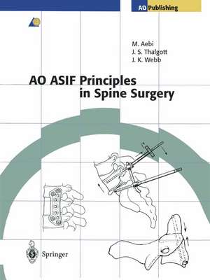
AO ASIF Principles in Spine Surgery
Contribuţii de M. Goytan Editat de Max Aebi, John S. Thalgott Contribuţii de B. Jeanneret, F. Magerl Editat de John K. Webb Contribuţii de M.B.Jr. Williamsonen Limba Engleză Paperback – 15 oct 2012
| Toate formatele și edițiile | Preț | Express |
|---|---|---|
| Paperback (1) | 1101.21 lei 6-8 săpt. | |
| Springer Berlin, Heidelberg – 15 oct 2012 | 1101.21 lei 6-8 săpt. | |
| Hardback (1) | 1289.69 lei 38-44 zile | |
| Springer Berlin, Heidelberg – 14 dec 1997 | 1289.69 lei 38-44 zile |
Preț: 1101.21 lei
Preț vechi: 1159.17 lei
-5% Nou
Puncte Express: 1652
Preț estimativ în valută:
210.78€ • 229.03$ • 177.17£
210.78€ • 229.03$ • 177.17£
Carte tipărită la comandă
Livrare economică 21 aprilie-05 mai
Preluare comenzi: 021 569.72.76
Specificații
ISBN-13: 9783642637469
ISBN-10: 3642637469
Pagini: 260
Ilustrații: XI, 243 p. 326 illus., 12 illus. in color.
Dimensiuni: 210 x 280 x 14 mm
Greutate: 0.59 kg
Ediția:Softcover reprint of the original 1st ed. 1998
Editura: Springer Berlin, Heidelberg
Colecția Springer
Locul publicării:Berlin, Heidelberg, Germany
ISBN-10: 3642637469
Pagini: 260
Ilustrații: XI, 243 p. 326 illus., 12 illus. in color.
Dimensiuni: 210 x 280 x 14 mm
Greutate: 0.59 kg
Ediția:Softcover reprint of the original 1st ed. 1998
Editura: Springer Berlin, Heidelberg
Colecția Springer
Locul publicării:Berlin, Heidelberg, Germany
Public țintă
Professional/practitionerDescriere
This book has become necessary as a consequence of the rapid expansion of the surgical procedures and implants available for spinal surgery within the "AO Group". We have not attempted to write an in-depth book on spinal surgery, but one which will help the surgeon in the use of AO concepts and implants. We con sider the practical courses held all over the world essential for the teaching of sound techniques so that technical complications and poor results can be avoid ed for both the surgeon and, in particular the patient. This book is a practical manual and an outline of what is taught in the courses. It is intended to help the young spinal surgeon to understand the correct use of AO implants. The indi- tions given will aid the correct use of each procedure. . It must be strongly emphasized that surgery of the spine is technically de manding. The techniques described in this book should only be undertaken by surgeons who are trained and experienced in spinal surgery. Certain techniques, in particular pedicle screw fIxation and cages, have not yet been fully approved by the FDA in the United States. However, throughout the rest of the world, the use of pedicle screws has become a standard technique for the spine surgeon, since it has been shown to improve fIxation techniques and allow segmental correction of the spine. The use of cages has become more and more popular, specifIcally as a tool of minimally invasive spinal surgery.
Cuprins
1 Aims and Principles.- 1.1 Introduction.- 1.2 Stable Internal Fixation.- 1.3 Preservation of Blood Supply.- 1.4 Anatomical Alignment.- 1.5 Early Pain-Free Mobilization.- 2 Biomechanics of the Spine and Spinal Instrumentation.- 2.1 Introduction.- 2.2 Mechanical Principles.- 2.3 Mechanical Properties of Materials.- 2.4 Implant Materials.- 2.5 Principles of Surgical Stabilization.- 2.5.1 Buttressing Principle.- 2.5.2 Neutralization Principle.- 2.5.3 Tension Band Principle.- 2.5.4 Bridge Fixation Principle.- 2.6 Instrumentation Application.- 2.7 Cervical Spinal Instrumentation.- 2.8 Lumbar and Thoracolumbar Spinal Instrumentation.- 2.9 Spinal Deformity.- 2.10 Lumbar Reconstruction.- 2.11 Spondylolysis.- 2.12 Implant Failure - Biomechanics.- References.- 3 Biology of Spinal Fusions.- 3.1 Introduction.- 3.2 Local Host Factors.- 3.2.1 Host Soft Tissue Bed.- 3.2.2 Host Graft Recipient Site.- 3.2.3 Growth Factors.- 3.2.4 Electrical Stimulation.- 3.3 Sytemic Host Factors.- 3.3.1 Hormones.- 3.3.2 Host Nutrition and Homeostasis.- 3.4 Bone Graft Materials.- 3.4.1 Properties of Graft Material.- 3.5 Types of Graft Materials.- 3.5.1 Autograft.- 3.5.2 Allograft.- 3.5.3 Xenograft.- 3.5.4 Synthetic Bone Graft Substitutes.- 3.5.5 Effect of Instrumentation on the Biology of Spine Fusions.- 3.6 Conclusion.- References.- 4 A Comprehensive Classification of Thoracic and Lumbar Injuries.- 4.1 Introduction.- 4.2 Concept of the Classification.- 4.3 Classification of Thoracic and Lumbar Injuries.- 4.3.1 Type A: Vertebral Body Compression.- 4.3.1.1 Group A1: Impaction Fractures.- 4.3.1.2 Group A2: Spilt Fractures.- 4.3.1.3 Group A3: Burst Fractures.- 4.3.1.4 Common Local Clinical Findings and Radiological Signs of Type A Injuries.- 4.3.2 Type B: Anterior and Posterior Element Injuries with Distraction.- 4.3.2.1 Group B1: Posterior Disruption Predominantly Ligamentous.- 4.3.2.2 Group B2: Posterior Disruption Predominantly Osseous.- 4.3.2.3 Common Local Clinical Findings and Radiological Signs of B1 and B2 Injuries.- 4.3.2.4 Group B3: Anterior Disruption Through the Disk.- 4.3.2.5 Common Local Clinical Findings and Radiological Signs of B3 Injuries.- 4.3.3 Type C: Anterior and Posterior Element Injuries with Rotation.- 4.3.3.1 Group Cl: Type A with Rotation.- 4.3.3.2 Group C2: Type B with Rotation.- 4.3.3.3 Group C3: Rotational Shear Injuries.- 4.3.3.4 Common Clinical Findings and Radiological Signs of Type C Injuries.- 4.4 Epidemiological Data.- 4.4.1 Level of Main Injury.- 4.4.2 Frequency and Distribution of Types and Groups.- 4.4.3 Incidence of Neurological Deficit.- 4.5 Discussion.- 4.5.1 Type A.- 4.5.2 Type B.- 4.5.2.1 B1 and B2 Injuries.- 4.5.2.2 B3 Injuries.- 4.5.3 Type C.- 4.6 Conclusions.- References.- 5 Stabilization Techniques: Upper Cervical Spine.- 5.1 Posterior Wiring Techniques.- 5.1.1 Standard Technique (After Gallie).- 5.1.2 Wedge Compression Technique (After Brooks and Jenkins).- 5.2 Transarticular Screw Fixation.- 5.2.1 Standard Technique.- 5.2.2 Cannulated Screw Technique.- 5.3 Anterior Screw Fixation of Odontoid (Dens) Fractures.- 5.3.1 Standard Lag Screw Technique.- 5.3.2 Cannulated Screw Technique.- 6 Stabilization Techniques: Lower Cervical Spine.- 6.1 Posterior Techniques.- 6.1.1 Wiring Technique.- 6.1.2 Plate Technique.- 6.1.2.1 Screw Placement.- 6.1.2.1.1 Mid and Lower Cervical Spine.- 6.1.2.1.2 Upper Cervical Spine.- 6.1.2.1.3 Upper Thoracic Spine from T1 - T3.- 6.1.2.1.4 Occiput Screws.- 6.1.2.2 3.5-mm Cervical Titanium Plate.- 6.1.2.2.1 Plate Fixation in the Middle and Lower Cervical Vertebrae (C2-C7).- 6.1.2.2.2 Occipitocervical Plate Fixation.- 6.1.2.2.3 Cervicothoracic Plate Fixation.- 6.1.2.3 One-Third Tubular Plate Fixation.- 6.1.2.4 Hook Plates.- 6.1.3 Cervical Spine Titanium Rod System (Cervifix).- 6.1.3.1 Implants and Instruments.- 6.1.3.2 Occipitocervical Stabilization.- 6.1.3.3 Cervicothoracic Fixation from C2 to Th2 (as for Occipitocervical Fixation).- 6.1.3.4 Connection of the Cervical Spine Rod System to the 6-mm USS Rod (Occipitocervical Fixation).- 6.2 Anterior Techniques.- 6.2.1 Plating Techniques.- 6.2.1.1 Standard H Plate.- 6.2.1.2 Titanium Cervical Spine Locking Plate (CSLP).- 7 Stabilization Techniques: Thoracolumbar Spine.- 7.1 Anterior Techniques.- 7.1.1 Plate Techniques.- 7.1.1.1 Fixation with the Large Dynamic Compression Plate (DCP).- 7.1.1.2 Anterior Titanium Thoracolumbar Locking Plate.- 7.1.2 Rod Systems.- 7.1.2.1 Fixation with the Anterior USS.- 7.1.2.1.1 Anterior Construct.- 7.1.2.1.2 Anterior Vertebral Body Construct.- 7.1.2.2 Fixation with the Anterior Titanium Rod System (VentroFix).- 7.1.2.2.1 Double-Rod Clamp Configuration.- 7.1.2.2.2 Fracture Clamp Configuration.- 7.1.2.2.3 Single-Clamp — Double-Rod Configuration.- 7.1.2.2.4 Single-Clamp — Single-Rod Configuration.- 7.2 Posterior Techniques.- 7.2.1 Translaminar Screw Fixation.- 7.2.2 Pedicle Fixation.- 7.2.2.1 Notched Thoracolumbar Plates.- 7.2.2.2 Fracture Module of the USS.- 7.2.2.2.1 Anterior Vertebral Body Fracture with Intact Posterior Wall (Type A-1 and A-2).- 7.2.2.2.2 Anterior Vertebral Body Fracture with Fractured Posterior Wall (Type A-3).- 7.2.2.2.3 Posterior Element Fractures or Disruption with Distraction (Type B).- 7.2.2.2.4 Complete Disruption of the Anterior and Posterior Elements with Rotation (Type C).- 8 Modular Stabilization System: The Universal Spine System.- 8.1 Basic Concepts.- 8.2 The System.- 8.2.1 Instruments and Implants.- 8.2.1.1 Fracture Module.- 8.2.1.2 Low Back Surgery Module.- 8.2.1.3 Scoliosis and Deformity Module.- 8.2.1.4 Specific Implants and Instruments.- 8.2.1.4.1 USS Side-Opening Pedicle Screws.- 8.2.1.4.2 USS Hooks: Laminar Hooks.- 8.2.1.4.3 USS Hooks: Specialized Pedicle Hook.- 8.2.1.4.4 USS Hooks: Transverse Process Hook.- 8.2.1.4.5 USS Rod Introduction into Side-Opening Implants.- 8.2.1.4.6 Complex Reduction Forceps — the “Persuader”.- 8.2.1.4.7 Rod Connectors.- 8.2.1.4.8 USS Cross-link System.- 8.2.2 USS for Deformity.- 8.2.2.1 Basic Principles.- 8.2.2.1.1 Basic Principle of Construct.- 8.2.2.1.2 The Concave Curve.- 8.2.2.1.3 The Convex Side.- 8.2.2.1.4 Insertion of Rod and Reduction of Spine.- 8.2.2.2 Scoliosis: Postrior Correction and Stabilization.- 8.2.2.2.1 Right Thoracic Late Onset Scoliosis.- 8.2.2.2.2 Double Curve.- 8.2.2.3 Scoliosis: Anterior Correction and Stabilization.- 8.2.2.4 Kyphosis: Posterior Correction and Stabilization.- 8.2.3 USS for the Degenerative Lumbosacral Spine.- 8.2.3.1 Standard Fixation.- 8.2.3.2 Sacral Fixation.- 8.2.4 USS for Spondylolisthesis Reduction and Stabilization.- 8.2.4.1 Spondylolisthesis.- 9 Other Fixation Systems.- 9.1 Hook-Screw System for Spondylolysis Treatment.- 9.2 External Spinal Skeletal Fixation B. Jeanneret.- 9.2.1 Principles and Technique.- 9.2.1.1 Technique of Percutaneous Insertion of Schanz Screws.- 9.2.1.2 Postoperative Care.- 9.2.1.3 Insertion of Thoracic Screws.- 9.2.1.4 Treatment of Complications.- 9.2.1.5 Removal of Schanz Screws.- 9.2.2 External Fixation as a Diagnostic Tool in Low-Back Pain.- 9.2.2.1 Gradual Reduction of Severe Spondylolisthesis.- 9.2.3 Percutaneous Treatment of Osteomyelitis of the Spine.- 9.2.4 External Fixation for Spinal Fractures.- 9.2.5 Stabilization of Unstable Malgaigne Fractures.- 9.3 Cage Systems.- 9.3.1 Fixation with the Titanium Interbody Spacer.- 9.3.2 Anterior Titanium Interbody Spacer (SynCage).- 9.3.3 Contact Fusion Cage.- 9.3.2.1 Mounting the Cage on the Implant Holder.- 9.3.2.2 Filling the Cage with Bone Graft.- 9.3.3.3 Removal of the Cage.
