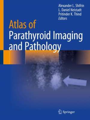
Atlas of Parathyroid Imaging and Pathology
Editat de Alexander L. Shifrin, L. Daniel Neistadt, Pritinder K. Thinden Limba Engleză Paperback – 7 oct 2021
This book provides a visual demonstration of normal and ectopic locations of parathyroid adenomas using different modalities in patients with PHPT and to describe parathyroid gland–related pathology. It includes several modern imaging modalities for localization of parathyroid glands and parathyroid adenomas, such as Sestamibi scan, SPECT/CT Sestamibi scan, neck ultrasound, MRI, thin-cut CT, and 4D CT scans. Written by experts in the field, chapters include pathology images corresponding to radiology imaging for some presented cases (gross and high-power view). Authors have also collected radiological images of difficult-to-localize parathyroid adenomas in ectopic (abnormal) locations. The atlas is organized by location of the adenomas in upper and lower eutopic locations followed by ectopic locations. Each case demonstrates dual or triple modalities such as US, Sestamibi scan, or SPECT/CT Sestamibi scan, thin-cut CT scan, or 4D CT performed on the same patient. A chapter on parathyroid pathology is also included to help the reader understand challenges in pathological interpretation.
Atlas of Parathyroid Imaging and Pathology serves as a valuable reference for radiologists, endocrine surgeons, head and neck surgeons, ENT surgeons, surgical oncologists, endocrinologists, pathologists, nephrologists, students, and all physicians and allayed health practitioners involved in the treatment of patients with primary, secondary, and tertiary hyperparathyroidism.
Atlas of Parathyroid Imaging and Pathology serves as a valuable reference for radiologists, endocrine surgeons, head and neck surgeons, ENT surgeons, surgical oncologists, endocrinologists, pathologists, nephrologists, students, and all physicians and allayed health practitioners involved in the treatment of patients with primary, secondary, and tertiary hyperparathyroidism.
| Toate formatele și edițiile | Preț | Express |
|---|---|---|
| Paperback (1) | 465.59 lei 38-44 zile | |
| Springer International Publishing – 7 oct 2021 | 465.59 lei 38-44 zile | |
| Hardback (1) | 637.42 lei 38-44 zile | |
| Springer International Publishing – 6 oct 2020 | 637.42 lei 38-44 zile |
Preț: 465.59 lei
Preț vechi: 490.10 lei
-5% Nou
Puncte Express: 698
Preț estimativ în valută:
89.10€ • 96.75$ • 74.84£
89.10€ • 96.75$ • 74.84£
Carte tipărită la comandă
Livrare economică 18-24 aprilie
Preluare comenzi: 021 569.72.76
Specificații
ISBN-13: 9783030409616
ISBN-10: 3030409619
Pagini: 283
Ilustrații: XIII, 283 p. 207 illus., 199 illus. in color.
Dimensiuni: 210 x 279 mm
Greutate: 0.84 kg
Ediția:2020
Editura: Springer International Publishing
Colecția Springer
Locul publicării:Cham, Switzerland
ISBN-10: 3030409619
Pagini: 283
Ilustrații: XIII, 283 p. 207 illus., 199 illus. in color.
Dimensiuni: 210 x 279 mm
Greutate: 0.84 kg
Ediția:2020
Editura: Springer International Publishing
Colecția Springer
Locul publicării:Cham, Switzerland
Cuprins
Parathyroid Ultrasound.- Scintigraphic Parathyroid Imaging.- Pathology of the Parathyroid Glands.- Right Superior Parathyroid Adenoma.- Right Inferior Parathyroid Adenoma.- Left Superior Parathyroid Adenoma.- Left Inferior Parathyroid Adenoma.- Ultrasonography, Sestamibi Scan, and SPECT/CT Sestamibi Scan of an Intrathyroidal Parathyroid Adenoma and Cystic Parathyroid Adenoma.- Imaging of the Parathyroid Carcinoma.- Motivation for Imaging Studies.- Contrast CT Approach.- The CT Technique.- Individual CT Phases.- Sources of False Positive and False Negative Enlarged Parathyroid Glands.- Correlative Ultrasound.- Shape, Number, and Size of Parathyroids.- Location of Parathyroid Glands.- Multigland Disease.- Rare Parathyroid Presentations.- Illustrative Cases.- Summary: CT Scan of the Neck in the Evaluation of Parathyroid Glands.- Invasive Techniques for Parathyroid Localization.- MRI of the Parathyroid Glands.
Notă biografică
Alexander Shifrin
Jersey Shore University Medical Center,
Department of Surgery
Neptune, NJ
USA
L. Daniel Neistadt
Lenox Hill Radiology
New York, NYUSA
Pritinder K. Thind
Jersey Shore University Medical Center
Department of Radiology
Neptune, NJ
USA
Textul de pe ultima copertă
This book provides a visual demonstration of normal and ectopic locations of parathyroid adenomas using different modalities in patients with PHPT and to describe parathyroid gland–related pathology. It includes several modern imaging modalities for localization of parathyroid glands and parathyroid adenomas, such as Sestamibi scan, SPECT/CT Sestamibi scan, neck ultrasound, MRI, thin-cut CT, and 4D CT scans. Written by experts in the field, chapters include pathology images corresponding to radiology imaging for some presented cases (gross and high-power view). Authors have also collected radiological images of difficult-to-localize parathyroid adenomas in ectopic (abnormal) locations. The atlas is organized by location of the adenomas in upper and lower eutopic locations followed by ectopic locations. Each case demonstrates dual or triple modalities such as US, Sestamibi scan, or SPECT/CT Sestamibi scan, thin-cut CT scan, or 4D CT performed on the same patient. A chapter on parathyroid pathology is also included to help the reader understand challenges in pathological interpretation.Atlas of Parathyroid Imaging and Pathology serves as a valuable reference for radiologists, endocrine surgeons, head and neck surgeons, ENT surgeons, surgical oncologists, endocrinologists, pathologists, nephrologists, students, and all physicians and allayed health practitioners involved in the treatment of patients with primary, secondary, and tertiary hyperparathyroidism.
Caracteristici
Provides visual demonstration of typical and abnormal (ectopic) localizations of parathyroid adenomas Each chapter will have very short description of the case accompanied by several different radiological images for the same case Includes several modern imaging modalities for localization of parathyroid glands and parathyroid adenomas
