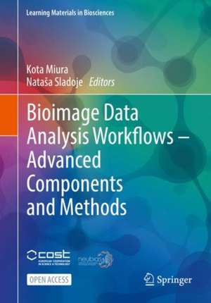
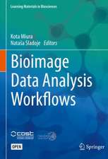
Bioimage Data Analysis Workflows ‒ Advanced Components and Methods: Learning Materials in Biosciences
Editat de Kota Miura, Nataša Sladojeen Limba Engleză Paperback – 30 sep 2022
Addressing the main challenges in image data analysis, where acquisition by powerful imaging devices results in very large amounts of collected image data, the book discusses techniques relying on batch and GPU programming, as well as on powerful deep learning-based algorithms. In addition, downstream data processing techniques are introduced, such as Python libraries for data organization, plotting, and visualizations. Finally, by studying the way individual unique ideas are implemented in the workflows, readers are carefully guided through how the parameters driving biological systems are revealed by analyzing image data. These studies include segmentation of plant tissue epidermis, analysis of the spatial pattern of the eye development in fruit flies, and the analysis of collective cell migration dynamics.
The presented content extends the Bioimage Data Analysis Workflows textbook (Miura, Sladoje, 2020), published in this same series, with new contributions and advanced material, while preserving the well-appreciated pedagogical approach adopted and promoted during the training schools for bioimage analysis organized within NEUBIAS – the Network of European Bioimage Analysts.
This textbook is intended for advanced students in various fields of the life sciences and biomedicine, as well as staff scientists and faculty members who conduct regular quantitative analyses of microscopy images.
Din seria Learning Materials in Biosciences
- 17%
 Preț: 394.87 lei
 - 20%
 Preț: 506.68 lei
 - 5%
 Preț: 529.76 lei
 - 5%
 Preț: 338.39 lei
 - 5%
 Preț: 330.25 lei
 -
 Preț: 347.49 lei
 - 5%
 Preț: 561.91 lei
 - 17%
 Preț: 426.67 lei
 - 17%
 Preț: 457.76 lei
 - 5%
 Preț: 491.25 lei
 - 17%
 Preț: 457.86 lei
 - 17%
 Preț: 490.61 lei
 -
 Preț: 358.43 lei
 - 17%
 Preț: 424.90 lei
 - 5%
 Preț: 414.83 lei
 - 17%
 Preț: 458.26 lei
 - 13%
 Preț: 383.66 lei
 - 17%
 Preț: 396.09 lei
 - 5%
 Preț: 797.65 lei
 - 20%
 Preț: 345.74 lei
 - 5%
 Preț: 797.65 lei
 - 17%
 Preț: 428.12 lei
 - 5%
 Preț: 671.48 lei
 - 19%
 Preț: 397.78 lei
 -
 Preț: 339.79 lei
 - 15%
 Preț: 527.97 lei
 - 19%
 Preț: 477.46 lei
 - 5%
 Preț: 583.96 lei
 - 19%
 Preț: 436.50 lei
 - 5%
 Preț: 527.90 lei
 - 15%
 Preț: 494.85 lei
 - 15%
 Preț: 466.45 lei
 - 15%
 Preț: 494.85 lei
 - 5%
 Preț: 524.97 lei
 - 15%
 Preț: 533.72 lei
 - 15%
 Preț: 528.99 lei
 - 5%
 Preț: 595.01 lei
 - 15%
 Preț: 596.19 lei
 - 15%
 Preț: 527.48 lei
Preț: 353.65 lei
Nou
Puncte Express: 530
Preț estimativ în valută:
67.68€ • 73.49$ • 56.85£
67.68€ • 73.49$ • 56.85£
Carte tipărită la comandă
Livrare economică 23 aprilie-07 mai
Preluare comenzi: 021 569.72.76
Specificații
ISBN-13: 9783030763930
ISBN-10: 3030763935
Pagini: 217
Ilustrații: X, 212 p. 265 illus. in color.
Dimensiuni: 168 x 240 x 17 mm
Greutate: 0.39 kg
Ediția:1st ed. 2022
Editura: Springer International Publishing
Colecția Springer
Seria Learning Materials in Biosciences
Locul publicării:Cham, Switzerland
ISBN-10: 3030763935
Pagini: 217
Ilustrații: X, 212 p. 265 illus. in color.
Dimensiuni: 168 x 240 x 17 mm
Greutate: 0.39 kg
Ediția:1st ed. 2022
Editura: Springer International Publishing
Colecția Springer
Seria Learning Materials in Biosciences
Locul publicării:Cham, Switzerland
Cuprins
Introduction.- Batch Processing Methods in ImageJ.- Python: Data Handling, Analysis and Plotting.- Building a Bioimage Analysis Workflow Using Deep Learning.- GPU-Accelerating ImageJ Macro Image Processing Workflows Using CLIJ.- How to Do the Deconstruction of Bioimage Analysis Workflows: A Case Study with SurfCut.- i.2.i. with the (Fruit) Fly: Quantifying Position Effect Variegation in Drosophila Melanogaster.- A MATLAB Pipeline for Spatiotemporal Quantification of Monolayer Cell Migration.
Notă biografică
Dr. Kota Miura is a Freelance Bioimage Analyst and works with various research groups and companies in Europe, for teaching, consulting, and collaborations. He also is affiliated with the Nikon Imaging Center at the University of Heidelberg and is the Vice-Chair of NEUBIAS (the Network of European Bioimage Analysts).
Nataša Sladoje is Professor in Computerized Image Processing at the Centre for Image Analysis, Department of Information Technology, Uppsala University, Sweden. She is coordinator of a Master’s programme in Image Analysis and Machine Learning at Uppsala University and has a long experience in education in this field. Her research interests include theoretical development of image analysis methods for robust image processing with high information preservation, robust methods for image comparison and registration, as well as development and applications of machine and deep learning methods particularly suitable for biomedical image analysis. She is the founder and leader of MIDA – Methods for Image Data Analysis – research group at Department of Information Technology, Uppsala University, and an active member of NEUBIAS – the European Network of Bioimage Analysis.
Nataša Sladoje is Professor in Computerized Image Processing at the Centre for Image Analysis, Department of Information Technology, Uppsala University, Sweden. She is coordinator of a Master’s programme in Image Analysis and Machine Learning at Uppsala University and has a long experience in education in this field. Her research interests include theoretical development of image analysis methods for robust image processing with high information preservation, robust methods for image comparison and registration, as well as development and applications of machine and deep learning methods particularly suitable for biomedical image analysis. She is the founder and leader of MIDA – Methods for Image Data Analysis – research group at Department of Information Technology, Uppsala University, and an active member of NEUBIAS – the European Network of Bioimage Analysis.
Textul de pe ultima copertă
This open access textbook aims at providing detailed explanations on how to design and construct image analysis workflows to successfully conduct bioimage analysis.
Addressing the main challenges in image data analysis, where acquisition by powerful imaging devices results in very large amounts of collected image data, the book discusses techniques relying on batch and GPU programming, as well as on powerful deep learning-based algorithms. In addition, downstream data processing techniques are introduced, such as Python libraries for data organization, plotting, and visualizations. Finally, by studying the way individual unique ideas are implemented in the workflows, readers are carefully guided through how the parameters driving biological systems are revealed by analyzing image data. These studies include segmentation of plant tissue epidermis, analysis of the spatial pattern of the eye development in fruit flies, and the analysis of collective cell migration dynamics.
The presented content extends the Bioimage Data Analysis Workflows textbook (Miura, Sladoje, 2020), published in this same series, with new contributions and advanced material, while preserving the well-appreciated pedagogical approach adopted and promoted during the training schools for bioimage analysis organized within NEUBIAS – the Network of European Bioimage Analysts.
This textbook is intended for advanced students in various fields of the life sciences and biomedicine, as well as staff scientists and faculty members who conduct regular quantitative analyses of microscopy images.
Caracteristici
This book is open access, which means that you have free and unlimited access The book explains computational methods in the context of biological questions Provides real examples and hands–on experience Gives detailed instructions on how to design and implement bioimage analysis workflow Builds upon the 1st volume “Bioimage Data Analysis Workflows” in this series Offers more advanced methods made understandable for a diverse group of users Meets the needs of this highly interdisciplinary field, which makes the content rather unique
