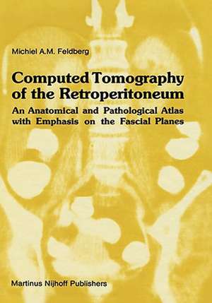
Computed Tomography of the Retroperitoneum: An Anatomical and Pathological Atlas with Emphasis on the Fascial Planes: Series in Radiology, cartea 8
Autor Michiel A.M. Feldbergen Limba Engleză Hardback – 30 sep 1983
| Toate formatele și edițiile | Preț | Express |
|---|---|---|
| Paperback (1) | 1397.27 lei 6-8 săpt. | |
| SPRINGER NETHERLANDS – 30 mar 2012 | 1397.27 lei 6-8 săpt. | |
| Hardback (1) | 1405.85 lei 6-8 săpt. | |
| SPRINGER NETHERLANDS – 30 sep 1983 | 1405.85 lei 6-8 săpt. |
Din seria Series in Radiology
- 5%
 Preț: 437.14 lei
 - 5%
 Preț: 367.51 lei
 - 5%
 Preț: 363.50 lei
 - 5%
 Preț: 348.95 lei
 - 5%
 Preț: 363.50 lei
 -
 Preț: 367.86 lei
 - 5%
 Preț: 366.74 lei
 - 5%
 Preț: 360.95 lei
 - 5%
 Preț: 393.79 lei
 - 5%
 Preț: 363.50 lei
 - 5%
 Preț: 340.64 lei
 - 5%
 Preț: 710.65 lei
 - 5%
 Preț: 704.85 lei
 - 5%
 Preț: 364.38 lei
 - 5%
 Preț: 710.65 lei
 - 5%
 Preț: 383.11 lei
 - 5%
 Preț: 1398.03 lei
 - 5%
 Preț: 376.91 lei
 - 5%
 Preț: 670.95 lei
Preț: 1405.85 lei
Preț vechi: 1479.84 lei
-5% Nou
Puncte Express: 2109
Preț estimativ în valută:
269.23€ • 274.74$ • 226.51£
269.23€ • 274.74$ • 226.51£
Carte tipărită la comandă
Livrare economică 26 februarie-12 martie
Preluare comenzi: 021 569.72.76
Specificații
ISBN-13: 9780898385731
ISBN-10: 0898385733
Pagini: 190
Ilustrații: XIV, 190 p.
Dimensiuni: 178 x 254 x 13 mm
Greutate: 0.58 kg
Ediția:1983
Editura: SPRINGER NETHERLANDS
Colecția Springer
Seria Series in Radiology
Locul publicării:Dordrecht, Netherlands
ISBN-10: 0898385733
Pagini: 190
Ilustrații: XIV, 190 p.
Dimensiuni: 178 x 254 x 13 mm
Greutate: 0.58 kg
Ediția:1983
Editura: SPRINGER NETHERLANDS
Colecția Springer
Seria Series in Radiology
Locul publicării:Dordrecht, Netherlands
Public țintă
ResearchCuprins
1. Case Material and Methods.- 1.1. Case materials.- 1.2. CT techniques.- 1.3. Patient preparation and contrast enhancement.- 2. Review of the literature. Anatomic considerations. Identification by CT.- 2.1. Introduction.- 2.2. History.- 2.3. Compartments of the retroperitoneum.- 2.4. Arrangements of the renal fascia.- 3. Gerota’s Fascia And Intraabdominal Fluid.- 3.1. Hemorrhage in retroperitoneum.- 3.2. Urinary extravasation in retroperitoneum.- 3.3. Acute and chronic inflammation of organs and structures in the retroperitoneal subspaces.- 3.4. Intraperitoneal fluid.- 4. Gerota’s Fascia And Infiltrating Malignancies.- 4.1. Primary retroperitoneal tumors.- 4.2. Renal cell carcinoma.- 4.3. Renal pelvis carcinoma.- 4.4. Wilms’ tumor (nephroblastoma).- 4.5. Adrenal tumors.- 4.6. Pancreatic tumor.- 4.7. Duodenum and ascending or descending colon tumor.- 5. Gerota’s Fascia Associated With LYMPH Node Disease of the Retrope- Ritoneum.- 5.1. General considerations.- 5.2. CT findings and illustrative cases.- Discussion of the Results.- Summary.- List of References.
Recenzii
`By mixing the text with superb CT images, Dr Feldberg has beautifully demonstrated this new dimension in retroperitoneal imaging. The extensive literature on the subject has been investigated and incorporated in the text. It is undoubtedly the most complete reference book on the subject. The study of this area - which in the past has been the domain reserved for the anatomist and surgeon - has now been opened to all. Because our studies are in the living patient, understanding of the spread of disease is now possible. Old and new techniques are combined and the result is a most thorough study of the retroperitoneal fascial planes.'
P.R. Koehler, Salt Lake City, UT, USA (from the Foreword)
`The text is concise and contains pertinent observations from the author's experience and a review of the literature. The atlas is adequately illustrated. The figures are clearly marked with appropriate arrows and symbols to aid the reader in identifying the important anatomic and pathologic features shown on the CT (Computed tomography) scans. The strength of the atlas lies in the cross-sectional and direct coronal CT illustrations. These illustrations provide a good method of studying the anatomy and pathology of the retroperitoneum and they also serve as an excellent reference.'
Journal of Computer Assisted Tomography, June 1984
