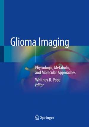
Glioma Imaging: Physiologic, Metabolic, and Molecular Approaches
Editat de Whitney B. Popeen Limba Engleză Paperback – 21 noi 2020
The text outlines current clinical practice including common scenarios in which imaging interpretation impacts patient management. Gaps in knowledge and potential areas of advancement based on the application of more experimental imaging techniques will be discussed. In reviewing this book, readers will learn:
- current standard imaging methodologies used in clinical practice for patients undergoing treatment for glioma and the implications of emerging treatment modalities including immunotherapy
- the theoretical basis for advanced imaging techniques including diffusion and perfusion MRI, MR spectroscopy, CEST and amino acid PET
- the relationship between imaging and molecular/genomic glioma features incorporated in the WHO 2016 classification update and the potential application of machine learning
- about the recently adopted and FDA approved standard brain tumor protocol for multicenter drug trials
- of the gaps in knowledge that impede optimal patient management and the cutting edge imaging techniques that could address these deficits
| Toate formatele și edițiile | Preț | Express |
|---|---|---|
| Paperback (1) | 563.96 lei 38-44 zile | |
| Springer International Publishing – 21 noi 2020 | 563.96 lei 38-44 zile | |
| Hardback (1) | 791.42 lei 38-44 zile | |
| Springer International Publishing – 21 noi 2019 | 791.42 lei 38-44 zile |
Preț: 563.96 lei
Preț vechi: 593.64 lei
-5% Nou
Puncte Express: 846
Preț estimativ în valută:
107.95€ • 117.29$ • 90.73£
107.95€ • 117.29$ • 90.73£
Carte tipărită la comandă
Livrare economică 17-23 aprilie
Preluare comenzi: 021 569.72.76
Specificații
ISBN-13: 9783030273613
ISBN-10: 303027361X
Pagini: 286
Ilustrații: XIV, 286 p. 109 illus., 69 illus. in color.
Dimensiuni: 178 x 254 mm
Ediția:1st ed. 2020
Editura: Springer International Publishing
Colecția Springer
Locul publicării:Cham, Switzerland
ISBN-10: 303027361X
Pagini: 286
Ilustrații: XIV, 286 p. 109 illus., 69 illus. in color.
Dimensiuni: 178 x 254 mm
Ediția:1st ed. 2020
Editura: Springer International Publishing
Colecția Springer
Locul publicării:Cham, Switzerland
Cuprins
1. Indications and Limitations of Conventional Imaging – Current Clinical Practice in the Context of Standard Therapy.- 2. Surrogates for Disease Status – Contrast Enhancement Including Limitations of Pseudoprogression and Pseudoresponse.- 3. The Relationship Between Biological and Imaging Characteristics in Enhancing and Nonenhancing Glioma.- 4. Contrast-Enhanced T1-Weighted Digital Subtraction for Increased Lesion Conspicuity and Quantifying Treatment Response in Malignant Gliomas.- 5. Advanced Physiologic Imaging: Perfusion. Theory and Applications.- 6. Advanced Physiologic Imaging: Diffusion. Theory and Applications.- 7. Parametric Response Map (PRM) Analysis Improves Response Assessment in Gliomas.- 8. Review of WHO 2016 Changes to Classification of Gliomas; Incorporation of Molecular Markers.- 9. Imaging markers of Low Grade Diffuse Glioma.- 10. CEST, pH and Glucose Imaging as Markers for Hypoxia and Malignant Transformation.- 11. MRS for D-2HG Detection in IDH Mutant Glioma.- 12. C-13 Hyperpolarized MR Spectroscopy for Metabolic Imaging of Brain Tumors.- 13. FET and FDOPA PET Imaging in Glioma.- 14. Imaging Genomics.- 15. Radiomics and Machine Learning.- 16. Immunotherapy and Gliomas.- 17. The Path Forward –The Standardized Brain Tumor Imaging Protocol (BTIP) for Multicenter Trials.
Notă biografică
Whitney B. Pope, MD, PhD is Professor-in-Residence and Director of Brian Tumor Imaging in the Department of Radiological Sciences at the David Geffen School of Medicine at UCLA. He has served on the editorial board of several neurology and neuroradiology journals, including Neuro-Oncology. He has presented and published widely on brain tumor imaging.
Textul de pe ultima copertă
This book covers physiologic, metabolic and molecular imaging for gliomas. Gliomas are the most common primary brain tumors. Imaging is critical for glioma management because of its ability to noninvasively define the anatomic location and extent of disease. While conventional MRI is used to guide current treatments, multiple studies suggest molecular features of gliomas may be identified with noninvasive imaging, including physiologic MRI and amino acid positron emission tomography (PET). These advanced imaging techniques have the promise to help elucidate underlying tumor biology and provide important information that could be integrated into routine clinical practice.
The text outlines current clinical practice including common scenarios in which imaging interpretation impacts patient management. Gaps in knowledge and potential areas of advancement based on the application of more experimental imaging techniques will be discussed. In reviewing this book, readers will learn:
The text outlines current clinical practice including common scenarios in which imaging interpretation impacts patient management. Gaps in knowledge and potential areas of advancement based on the application of more experimental imaging techniques will be discussed. In reviewing this book, readers will learn:
- current standard imaging methodologies used in clinical practice for patients undergoing treatment for glioma and the implications of emerging treatment modalities including immunotherapy
- the theoretical basis for advanced imaging techniques including diffusion and perfusion MRI, MR spectroscopy, CEST and amino acid PET
- the relationship between imaging and molecular/genomic glioma features incorporated in the WHO 2016 classification update and the potential application of machine learning
- about the recently adopted and FDA approved standard brain tumor protocol for multicenter drug trials
- of the gaps in knowledge that impede optimal patient management and the cutting edge imaging techniques that could address these deficits
Caracteristici
Focuses on an unmet clinical need for gliomas, including early response and predictive imaging markers that have the potential to truly impact patient management decisions In depth coverage of advanced physiologic imaging, including CEST, pH imaging, hyperpolarized MR spectroscopy, and 2-HG detection (for IDH1 mutant tumors) with MRS Integrates imaging with the updated World Health Organization brain tumor classification system Includes exposition on the new FDA supported standardized brain tumor imaging protocol, required for imagers to participate in upcoming trials Covers of radiogenomics, radiomics, and machine learning, cutting edge “big data” approaches to improving imaging for cancer and other diseases
