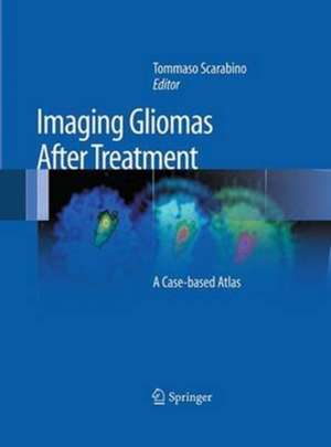
Imaging Gliomas After Treatment: A Case-based Atlas
Editat de Tommaso Scarabinoen Limba Engleză Paperback – 23 aug 2016
| Toate formatele și edițiile | Preț | Express |
|---|---|---|
| Paperback (1) | 648.03 lei 38-44 zile | |
| Springer – 23 aug 2016 | 648.03 lei 38-44 zile | |
| Hardback (1) | 923.19 lei 38-44 zile | |
| Springer – 21 noi 2011 | 923.19 lei 38-44 zile |
Preț: 648.03 lei
Preț vechi: 682.13 lei
-5% Nou
Puncte Express: 972
Preț estimativ în valută:
124.01€ • 134.66$ • 104.17£
124.01€ • 134.66$ • 104.17£
Carte tipărită la comandă
Livrare economică 19-25 aprilie
Preluare comenzi: 021 569.72.76
Specificații
ISBN-13: 9788847058262
ISBN-10: 8847058260
Pagini: 201
Ilustrații: XVIII, 201 p.
Dimensiuni: 193 x 260 mm
Ediția:Softcover reprint of the original 1st ed. 2012
Editura: Springer
Colecția Springer
Locul publicării:Milano, Italy
ISBN-10: 8847058260
Pagini: 201
Ilustrații: XVIII, 201 p.
Dimensiuni: 193 x 260 mm
Ediția:Softcover reprint of the original 1st ed. 2012
Editura: Springer
Colecția Springer
Locul publicării:Milano, Italy
Cuprins
Introduction.- Classification.- Gliomas.- Etiology – Heredity – Risk factors – Pathogenesis – Prognostic factors.- Complications – Signs and symptoms – Diagnosis and follow-up – Supportive therapy.- Treatment of gliomas.- Surgery – Radiotherapy – Chemotherapy.- Post-treatment neuroradiological imaging.-Morphological magnetic resonance.- Functional magnetic resonance: Spectroscopy – Diffusion – Perfusion – Cortical activation.- Clinical cases.- Bibliography.
Textul de pe ultima copertă
Patients with gliomas frequently undergo combination therapy that can result in complex and potentially confusing appearances on follow-up imaging. In this context, differentiation between tumor recurrence and radiation necrosis, for example, can be very difficult.
This atlas is a detailed guide to the imaging appearances of gliomas following treatment with neurosurgery, radiation therapy, and chemotherapy. Normal and pathological findings are displayed in detailed MR images that illustrate the potential modifications due to treatment. Particular emphasis is placed on characteristic appearances on the newer functional MR imaging techniques, including MR spectroscopy, diffusion-weighted imaging, and perfusion imaging. These techniques are revolutionizing neuroradiology by going beyond the demonstration of macroscopic alterations to the depiction of preceding metabolic changes at the cellular and subcellular level. They thereby improve the accuracy, sensitivity, and specificity of MR imaging and allow earlier and more specific diagnosis. A key section comprising some 40 clinical cases and more than 500 illustrations offers an invaluable clinical and research tool not only for neuroradiologists but also for neurosurgeons, radiotherapists, and medical oncologists.
This atlas is a detailed guide to the imaging appearances of gliomas following treatment with neurosurgery, radiation therapy, and chemotherapy. Normal and pathological findings are displayed in detailed MR images that illustrate the potential modifications due to treatment. Particular emphasis is placed on characteristic appearances on the newer functional MR imaging techniques, including MR spectroscopy, diffusion-weighted imaging, and perfusion imaging. These techniques are revolutionizing neuroradiology by going beyond the demonstration of macroscopic alterations to the depiction of preceding metabolic changes at the cellular and subcellular level. They thereby improve the accuracy, sensitivity, and specificity of MR imaging and allow earlier and more specific diagnosis. A key section comprising some 40 clinical cases and more than 500 illustrations offers an invaluable clinical and research tool not only for neuroradiologists but also for neurosurgeons, radiotherapists, and medical oncologists.
Caracteristici
First atlas devoted to the imaging appearances of gliomas following treatment Special attention to the role of the newer functional MR imaging techniques Includes 40 clinical cases with more than 500 illustrations, making the book an invaluable resource for everyday practice and research
Descriere
Descriere de la o altă ediție sau format:
This atlas is a detailed guide to the imaging appearances of gliomas following treatment with neurosurgery, radiation therapy and chemotherapy. It features more than 40 clinical cases and 500 illustrations, making it an invaluable clinical and research tool.
This atlas is a detailed guide to the imaging appearances of gliomas following treatment with neurosurgery, radiation therapy and chemotherapy. It features more than 40 clinical cases and 500 illustrations, making it an invaluable clinical and research tool.
