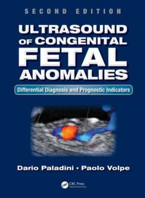
Ultrasound of Congenital Fetal Anomalies: Differential Diagnosis and Prognostic Indicators, Second Edition
Autor Dario Paladini, Paolo Volpeen Limba Engleză Hardback – 4 iun 2014
| Toate formatele și edițiile | Preț | Express |
|---|---|---|
| Hardback (2) | 1565.91 lei 6-8 săpt. | +276.76 lei 7-13 zile |
| CRC Press – 4 iun 2014 | 2163.91 lei 3-5 săpt. | +45.51 lei 7-13 zile |
| CRC Press – 14 iun 2024 | 1565.91 lei 6-8 săpt. | +276.76 lei 7-13 zile |
Preț: 2163.91 lei
Preț vechi: 2277.81 lei
-5% Nou
Puncte Express: 3246
Preț estimativ în valută:
414.05€ • 432.31$ • 342.69£
414.05€ • 432.31$ • 342.69£
Carte disponibilă
Livrare economică 14-28 martie
Livrare express 28 februarie-06 martie pentru 55.50 lei
Preluare comenzi: 021 569.72.76
Specificații
ISBN-13: 9781466598966
ISBN-10: 1466598964
Pagini: 516
Ilustrații: 480 black & white illustrations, 68 black & white tables
Dimensiuni: 210 x 280 x 25 mm
Greutate: 1.11 kg
Ediția:Revizuită
Editura: CRC Press
Colecția CRC Press
Locul publicării:Boca Raton, United States
ISBN-10: 1466598964
Pagini: 516
Ilustrații: 480 black & white illustrations, 68 black & white tables
Dimensiuni: 210 x 280 x 25 mm
Greutate: 1.11 kg
Ediția:Revizuită
Editura: CRC Press
Colecția CRC Press
Locul publicării:Boca Raton, United States
Public țintă
Professional ReferenceCuprins
Anatomic Survey of the Fetus and Early Diagnosis of Fetal Anomalies. Central and Peripheral Nervous System Anomalies. Craniofacial and Neck Anomalies. Cystic Hygroma and Nonimmune Hydrops Fetalis. Congenital Heart Disease. Thoracic Anomalies. Anomalies of the Gastrointestinal Tract and of the Abdominal Wall. Anomalies of the Urinary Tract and of the External Genitalia. Skeletal Dysplasias and Muscular Anomalies: A Diagnostic Algorithm. Chromosomal and Nonchromosomal Syndromes. Ultrasound in Fetal Infection. Ultrasound in Multiple Pregnancy. Appendix.
Recenzii
"In summary, this is a beautifully illustrated book which usefully presents US images, line diagrams, and clinical photographs which will enable the reader to better identify anatomical abnormalities."
—RAD Magazine
"This is a solid book with good images and some interesting features… [It] will be helpful for clinicians and sonographers who specialize in the care of complicated obstetrical patients."
—Anthony Shanks, MD, Washington University School of Medicine, in Doody's Book Review Service
"… an invaluable help in the ultrasound room, as well as an initial source for a thorough investigation of any particular anomaly or risk factor. … The extensive use of illustrated flowcharts to highlight differential diagnoses, as well as identification of the abnormalities from specific ultrasound features, will become significant assets in prenatal diagnosis. Another strong point of this volume is the continuous effort to include 3D images for most diagnoses, especially those that are most likely to benefit from this development, including cranial sutures, skeleton, and fetal brain and heart. … this new edition of Ultrasound of Congenital Fetal Anomalies, already an exhaustive reference with a unique pedagogic format, will become an important landmark in the field and will smoothly reach classic status on the way to its third edition!"
—From the Foreword by Yves Ville, Service of Obstetrics–Gynecology Necker-Enfants-Malades Hospital, Paris, France
Descriere
This new edition of an acclaimed text guides readers through the use of ultrasound to detect and identify a wide range of birth defects in utero. Ultrasound images have been improved throughout and are now matched with MRI scans as a further means of guiding the clinician. As further correlation, clinical images are also provided. This new edition also extends its approach to the fetus in the first trimester.
Notă biografică
Dario Paladini completed his internship at the University Medical school in Naples, Italy. He completed his residency in Obstetrics and Gynecology in 1990 at the Federico II University of Naples. In 1990-94 he moved to the National Cancer Institute of Milan for training in Gynecologic Oncology; he then returned to Naples and joined the faculty as lecturer and, since 2001, as associate professor of obstetrics and gynecology.
Medicina Fetale e Diagnosi Prenatale
Paolo Volpe is Director of Obstetrics and Gynecology at Ospedale "Di Venere", Carbonara di Bari, Italy.
Medicina Fetale e Diagnosi Prenatale
Paolo Volpe is Director of Obstetrics and Gynecology at Ospedale "Di Venere", Carbonara di Bari, Italy.
