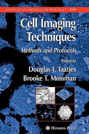
Cell Imaging Techniques: Methods in Molecular Biology, cartea 319
Editat de Douglas J. Taatjes, Brooke T. Mossmanen Limba Engleză Paperback – 10 noi 2010
Din seria Methods in Molecular Biology
- 9%
 Preț: 791.59 lei
 - 23%
 Preț: 598.56 lei
 - 20%
 Preț: 882.95 lei
 -
 Preț: 252.04 lei
 - 5%
 Preț: 802.69 lei
 - 5%
 Preț: 729.61 lei
 - 5%
 Preț: 731.43 lei
 - 5%
 Preț: 741.30 lei
 - 5%
 Preț: 747.16 lei
 - 15%
 Preț: 663.45 lei
 - 18%
 Preț: 1025.34 lei
 - 5%
 Preț: 734.57 lei
 - 18%
 Preț: 914.20 lei
 - 15%
 Preț: 664.61 lei
 - 15%
 Preț: 654.12 lei
 - 18%
 Preț: 1414.74 lei
 - 5%
 Preț: 742.60 lei
 - 20%
 Preț: 821.63 lei
 - 18%
 Preț: 972.30 lei
 - 15%
 Preț: 660.49 lei
 - 5%
 Preț: 738.41 lei
 - 18%
 Preț: 984.92 lei
 - 5%
 Preț: 733.29 lei
 -
 Preț: 392.58 lei
 - 5%
 Preț: 746.26 lei
 - 18%
 Preț: 962.66 lei
 - 23%
 Preț: 860.21 lei
 - 15%
 Preț: 652.64 lei
 - 5%
 Preț: 1055.50 lei
 - 23%
 Preț: 883.85 lei
 - 19%
 Preț: 491.88 lei
 - 5%
 Preț: 1038.84 lei
 - 5%
 Preț: 524.15 lei
 - 18%
 Preț: 2122.34 lei
 - 5%
 Preț: 1299.23 lei
 - 5%
 Preț: 1339.10 lei
 - 18%
 Preț: 1390.26 lei
 - 18%
 Preț: 1395.63 lei
 - 18%
 Preț: 1129.65 lei
 - 18%
 Preț: 1408.26 lei
 - 18%
 Preț: 1124.92 lei
 - 18%
 Preț: 966.27 lei
 - 5%
 Preț: 1299.99 lei
 - 5%
 Preț: 1108.51 lei
 - 5%
 Preț: 983.72 lei
 - 5%
 Preț: 728.16 lei
 - 18%
 Preț: 1118.62 lei
 - 18%
 Preț: 955.25 lei
 - 5%
 Preț: 1035.60 lei
 - 18%
 Preț: 1400.35 lei
Preț: 1290.07 lei
Preț vechi: 1357.98 lei
-5% Nou
Puncte Express: 1935
Preț estimativ în valută:
246.85€ • 258.43$ • 204.26£
246.85€ • 258.43$ • 204.26£
Carte tipărită la comandă
Livrare economică 05-19 aprilie
Preluare comenzi: 021 569.72.76
Specificații
ISBN-13: 9781617373947
ISBN-10: 161737394X
Pagini: 490
Ilustrații: XIV, 490 p. 212 illus.
Dimensiuni: 152 x 229 x 29 mm
Greutate: 0.74 kg
Ediția:Softcover reprint of hardcover 1st ed. 2006
Editura: Humana Press Inc.
Colecția Humana
Seria Methods in Molecular Biology
Locul publicării:Totowa, NJ, United States
ISBN-10: 161737394X
Pagini: 490
Ilustrații: XIV, 490 p. 212 illus.
Dimensiuni: 152 x 229 x 29 mm
Greutate: 0.74 kg
Ediția:Softcover reprint of hardcover 1st ed. 2006
Editura: Humana Press Inc.
Colecția Humana
Seria Methods in Molecular Biology
Locul publicării:Totowa, NJ, United States
Public țintă
ResearchCuprins
Molecular Beacons.- Second-Harmonic Imaging of Collagen.- Visualizing Calcium Signaling in Cells by Digitized Wide-Field and Confocal Fluorescent Microscopy.- Multifluorescence Labeling Techniques and Confocal Laser Scanning Microscopy on Lung Tissue.- Evaluation of Confocal Microscopy System Performance.- Quantitative Analysis of Atherosclerotic Lesion Composition in Mice.- Applications of Microscopy to Genetic Therapy of Cystic Fibrosis and Other Human Diseases.- Laser Scanning Cytometry.- Near-Clinical Applications of Laser Scanning Cytometry.- Laser Capture Microdissection.- Analysis of Asbestos-Induced Gene Expression Changes in Bronchiolar Epithelial Cells Using Laser Capture Microdissection and Quantitative Reverse Transcriptase-Polymerase Chain Reaction.- New Approaches to Fluorescence In Situ Hybridization.- Microarray Image Scanning.- Near-Field Scanning Optical Microscopy in Cell Biology and Cytogenetics.- Porosome.- Secretory Vesicle Swelling by Atomic Force Microscopy.- Imaging and Probing Cell Mechanical Properties With the Atomic Force Microscope.- Reflection Contrast Microscopy.- Three-Dimensional Analysis of Single Particles by Electron Microscopy.- Three-Dimensional Reconstruction of Single Particles in Electron Microscopy.- A New Microbiopsy System Enables Rapid Preparation of Tissue for High-Pressure Freezing.
Recenzii
From the reviews:
"...deserves to do well." -Ultramicroscopy
"The book covers a wide range of experimental imaging methods for the investigation of cells. … The book is well produced, with many excellent photographs and diagrams, and I have enjoyed broadening my awareness of techniques that not only did I not know in detail but in some cases not even know existed. A nice book … ." (D. J. Cook, British Journal of Biomedical Science, Vol. 63 (2), 2006)
"This 21-chapter volume … provides detailed descriptions of and protocols for a plethora of microscopy and related methods to study tissues, cells and macromolecules. … This comprehensive and well rounded book is suitable for institutional or personal purchase, novices and experts, and it is certainly a must-read for all serious confocal microscopists and imaging facility managers." (Andreas Holzenburg, Microbiology Today, August, 2006)
"Douglas Taatjes and Brooke Mossman have edited a compilation of 21 articles. … The subjects covered are very diverse, ranging from light microscopy to electron microscopy. … In summary, this book provides an excellent overview on current and relevant imaging techniques. The described methods and protocols are not only of particular interest to those who are new in the field or who have to expand their imaging capability above regular light microscopy. The book will serve as a reference for years to come … ." (Carsten Schultz, ChemBioChem, Vol. 7, 2006)
"This text will be extremely useful. … it is a collection of advanced and sometimes disparate microscopy techniques and their application to biological research. … The authors include highly detailed descriptions and instructions on the low-level methods computer programmers use for acquisition, storage, and manipulation of digital image data." (Steven Ruzin, Microscope, Vol. 54, 2006)
Textul de pe ultima copertă
Cell imaging methodologies have now become essential research tools for a variety of disciplines that traditionally had not relied on them. In Cell Imaging Techniques: Methods and Protocols, distinguished international researchers describe in detail their state-of-the-art methods for the microscopic imaging of cells and molecules. The authors cover a wide spectrum of complementary techniques, including such methods as fluorescence microscopy, electron microscopy, atomic force microscopy, and laser scanning cytometry. Additional protocols on confocal scanning laser microscopy, quantitative computer-assisted image analysis, laser-capture microdissection, microarray image scanning, near-field scanning optical microscopy, and reflection contrast microscopy round out this eclectic collection of cutting-edge imaging techniques now available. The authors also discuss preparative methods for particles and cells by transmission electron microscopy. The protocols follow the successful Methods in Molecular Biology™ series format, each offering step-by-step laboratory instructions, an introduction outlining the principles behind the technique, lists of the necessary equipment and reagents, and tips on troubleshooting and avoiding known pitfalls.
Timely and highly practical, Cell Imaging Techniques: Methods and Protocols provides researchers and clinicians with a richly useful guide to selecting and performing the best imaging method from a bewildering variety of microscopy-based techniques.
Timely and highly practical, Cell Imaging Techniques: Methods and Protocols provides researchers and clinicians with a richly useful guide to selecting and performing the best imaging method from a bewildering variety of microscopy-based techniques.
Caracteristici
Includes supplementary material: sn.pub/extras
